
Created by - Rigomo Team
Anaphylaxis – Disease Symptoms Diagnosis & Management
Anaphylaxis (an-a-fi-LAK-sis) could be a serious, grievous form of allergy. The commonest cause includes common hypersensitivity reactions to foods, insect stings, medications, and latex.If you're allergic to a substance, your system overreacts to that particular substance causing allergic reaction symptoms. Typically, these vexatious symptoms occur in one location of the body. However, some folks are liable to a way additional serious hypersensitivity reaction. This reaction usually affects one part of the body at the same time.Anaphylaxis needs immediate medical treatment, including a prompt injection of Adrenalin and a visit to a hospital ER. If it isn’t treated properly and timely, a hypersensitivity reaction can be fatal.Certain folks are particularly at higher risk of serious hypersensitivity reactions. If you have a history of allergies or asthma attacks With a history of hypersensitivity reactionsAccurate identification and immediate management of allergies are important. Symptoms of hypersensitivity reaction usually begin within five to half-hour of returning into contact with the substance that you're allergic to. In some cases, immediate intervention is needed. Symptoms of anaphylaxis –Warning signs are usually on one part of the body and should include:• Red rash, with hives/welts, that's sometimes fidgety (It is feasible to have a severe allergy while no skin symptoms.)• Swollen throat or swollen areas of the body (It is feasible to have a severe allergy while no skin symptoms.)• Wheezing• Passing out/unconsciousness• Chest tightness• Trouble breathing Nagging cough• Hoarse voice• Trouble swallowing• Vomiting• Diarrhea• Stomach cramping• Feeling of impending doomDiagnosisTo diagnose your risk of hypersensitivity reaction (anaphylaxis) or to see whether or not previous symptoms were anaphylaxis-related, you can conduct a radical investigation of all potential allergens. Your Doctor can provide you with specific details concerning all past sensitivity.ManagementThe best ways to manage your condition are:• Avoid allergens that trigger your sensitivity• Be ready for an immediate visit to the emergency for timely interventionIf you're at high risk of getting a hypersensitivity reaction (anaphylaxis), carry Adrenalin injectors (adrenaline). They contain a prescribed single dose of medication that's injected into the thigh for hypersensitivity reaction during an emergency.Be sure to speak to your caretaker/partner/family member about a way to use the Adrenalin appliance. It is helpful if you keep a diary related to your allergic reactions (Anaphylaxis). Write an action plan, and keeps it with you at work, school, camp, or different places wherever others might have to acknowledge your symptoms and supply treatment.
More detailsPublished - Tue, 19 Jul 2022

Created by - Rigomo Team
ACUTE RESPIRATORY FAILURE
Acute respiratory failure is defined as an acute impairment in oxygen or carbon dioxide exchange that results in or has the potential to result in patient morbidity or mortality. Impaired gas exchange (e.g., as a result of shunting, alveolar hypoventilation, ventilation-perfusion mismatch, or decreased pulmonary diffusion capacity) leads to hypoxemia (decreased arterial oxygen tension [PaO2]) and possibly hypoxia (i.e., insufficient delivery of oxygen to tissues). Impaired ventilation causes hypercapnia (i.e., an elevated carbon dioxide tension [PCO2]). Because the baseline respiratory status may vary greatly among patients, it is difficult to characterize acute respiratory insufficiency by purely numeric criteria.Generally, a patient who acutely develops a PO2 below 60 mmHg on room air or a PCO2 greater than 50 mm Hg with an arterial blood pH less than 7.35 is considered to be in respiratory failure.Some patients (e.g., those with chronic obstructive pulmonary disease [COPD]) chronically have an elevated PCO2 but a normal arterial blood pH. In these patients, respiratory failure is defined as a PCO2 higher than baseline with a concurrent decrease in serum pH.Causes: There are many causes of acute respiratory failure, including most pulmonary diseases as well as many cardiac diseases. Most commonly, one should consider airway obstruction, asthma, COPD, congestive heart failure (CHF), non-cardiogenic pulmonary edema, pulmonary emboli, massive pleural effusion, pneumonia, hemothorax, pneumothorax, toxic inhalation, advanced lung cancer, and neurologic or muscular disorders that result in impaired respiratory abilities.Symptoms: Dyspnea is the most common symptom of acute respiratory failure. With severe hypoxia, confusion, agitation, and disorientation might happen. Patients with significant hypercapnia frequently exhibit sluggishness or somnolence. Generalized seizures or coma may result from profound central nervous system hypoxia. Other signs of respiratory failure include cyanosis, diaphoresis, and severely labored breathing (including the use of accessory muscles). Children may demonstrate nasal flaring, audible grunting, or retractions (i.e., intercostal, subcostal, suprasternal).Physical examination findingsMild hypertension, tachypnea, and tachycardia are frequently observed.Patients with hypercapnia may exhibit bradypnea. Wheezing, rales, or decreased breath sounds may be noted on pulmonary examination, depending on the underlying disease.Evaluation: In a patient with suspected acute respiratory failure, patient evaluation and treatment occur simultaneously. A brief, focused clinical history and physical examination, including vital signs, will often reveal the cause of respiratory failure before obtaining any diagnostic studies. Chest radiography helps determine the underlying cause of acute respiratory failure. At the patient's bedside, radiographs should be taken as soon as possible. An Arterial blood gas (ABG) should be obtained in all patients with suspected acute respiratory failure. The PO2, PCO2, and pH make up the ABG's most crucial components. The patient's oxygen saturation may be measured instantly using pulse oximetry. Ancillary tests may be indicated, depending on the cause of the respiratory failure (e.g., ABG with co-oximetry for dyshemoglobinemias, an electrocardiogram [ECG] for a patient with suspected CHF, a V/Q· scan or computed tomography [CT] pulmonary angiogram for a patient with suspected pulmonary embolism, a urine toxicology screen for a patient with suspected narcotic overdose).Therapy of Airway, breathing, circulation (ABC): Airway management, usually orotracheal intubation, is required for patients who do not have an intact airway or who are unable to protect their airway. After an airway has been established, ensuring adequate oxygenation and ventilation is the mainstay of treating respiratory failure. Patients should be administered supplemental oxygen to maintain a serum oxygen saturation of at least 90%. In addition to oxygenation, ventilation needs to be taken care of. Patients with a PCO2 greater than 50mmHg and an arterial blood pH below 7.30 require intubation and mechanical ventilation if their condition cannot be quickly improved.Following intubation, ventilator settings for respiratory rates and tidal volumes should generally be adjusted to gradually normalize the blood pH. In general, the initial respiratory rate should be 12 to 16 breaths/min, with a tidal volume of 6 to 8 mL/kg.Ventilation is typically less significant than oxygenation. For this reason, it is acceptable to have mild respiratory acidosis (e.g., a pH less than 7.35) if necessary to maintain adequate oxygenation or to minimize peak airway pressures.Circulation: The placement of two peripheral intravenous lines allows the administration of fluids and medications to the patient.Specific therapy that addresses the cause of acute respiratory failure should be undertaken after the ABCs have been addressed. In some cases, aggressive treatment of the underlying condition may reverse the respiratory failure and eliminate the need for intubation (e.g., nitroglycerin and afterload reduction for CHF, inhaled β2 agonists for asthma or COPD, chest tube placement for a large pneumothorax or hemothorax).Disposition: All patients with acute respiratory failure should be admitted to an intensive care unit (ICU). Patients who are clinically stable but have the potential for developing respiratory failure should be admitted to an ICU or another closely monitored unit.
More detailsPublished - Wed, 20 Jul 2022

Created by - Rigomo Team
Diabetes – Types, Disorder & Symptoms
Diabetes mellitus is a disorder in which the blood sugar levels are more than normal. PrediabetesPrediabetes is a condition within which blood sugar levels are high but not high enough to be considered Diabetes. Folks have prediabetes if their fasting blood sugar level is between 100mg/dL (5.6 mmol/L) and 125 mg/dL (6.9 mmol/L) or if their glucose tolerance levels is between 140 mg/dL (7.8 mmol/L) and 199 mg/dL (11.0 mmol/L). Prediabetes increases the risk of developing type 2 Diabetes. Decreasing weight by five to 100 percent through diet and exercise will considerably scale back the danger of developing the future disorder.Type-1 Diabetes In Type 1diabetes (formerly known as insulin-dependent or juvenile-onset diabetes), the body's immune system attacks the insulin-producing cells of the pancreas and consequently destroys them. The gland, therefore, produces very little or no Insulin. Scientists believe that environmental issues—possibly an infection or stressful event during childhood or early adulthood in a genetically prone individual may trigger the autoimmune process causing the system to destroy the insulin-producing cells. of the duct gland. Type-2 Diabetes Type 2 diabetes is a chronic disorder (formerly known as a non–insulin-dependent disorder or adult-onset diabetes), the duct gland typically produces insulin, However, the body develops resistance to whatever insulin is made, thus there's not enough insulin to satisfy the body’s needs. As the disease progresses, the insulin-producing ability of the pancreas decreases.This type of chronic type2 diabetes is rare in youngsters and adolescents however have now become common. However, it always begins in folks older than thirty and becomes more common with age. Almost twenty-sixth % of individuals older than sixty-five have type 2 Diabetes.Signs and Symptoms of Diabetes –› Increased thirst› Frequent urination› Extreme fatigue or tiredness› Mental status changes› Unexplained and rapid weight loss› Blurred vision› Nausea and vomiting› Increased Hunger› Recurrent infections› Poor healing of wounds› Skin problems› Tingling or numbness in toe /feet Diabetes damages blood vessels and will increase the danger of heart failure, stroke, chronic nephropathy, and vision loss.Diagnosis is done by elevated fasting plasma glucose level and/or elevated HbA1C by your physician. Diabetes can be effectively managed through medicines along with diet, exercise, and lifestyle modification.
More detailsPublished - Wed, 20 Jul 2022

Created by - Rigomo Team
Is Kasayam /Kada universal for all or must be customized?
Kashāya or Kada is a combination of spices cooked in water that helps build immunity. The drink provides you relief from cold and cough. It’s often believed to be a nasty-tasting, medicine.This COVID-19 pandemic has taught us that increasing immunity in a natural way is the best way as it saves us from infections and diseases. This has paved the way to incorporate natural and organic ingredients into the diet.Kashaya or kadha is one of the most effective drinks in India with a lot of health advantages. Daily consumption of this drink is usually recommended by the AYUSH [ that Yoga, Unani, Siddha, and homeopathy] during this pandemic state. of affairs. Though, over many years, this kadha formulation has been used effectively for numerous issues.It is a conventional, homemade, aromatic drink that has various healing properties. Different medicative ingredients are used to make this kadha. It’s been employed in India since an earlier period however presently; it has come into the limelight due to the corona pandemic. Aside from boosting your immunity, this kadha can help relax your body, detoxifies the body, improve skin quality, strengthen your digestive health, and etc.PreparationFew common kitchen ingredients can be used to make an effective kadha recipe such as Turmeric, cumin seeds, pepper, ginger, etc. As these ingredients are hot in potency, you add them to the drink as long as your cold and cough are there. There are several versions of tea within the market however there are some basic ingredients that you ought to grasp –Ingredients1 tsp – turmeric powder (haldi)1 tsp – Cummin seeds (jeera)5 to 6 – Black peppercornsA pinch of – Fennel seeds (saunf)1/4 cup – MilkOptional – Tulsi ( 5/6 leaves)Ginger powder (a pinch)Method of making Kashya or KadaCrush cumin seeds, peppercorns, saunf, and ginger using a mortar pestle into a rough powder.Boil this powder in a cup and half water with turmeric.Let it boil well until the spices offer out all the flavor.Add milk and produce it to boil only for one or two seconds.Strain it and serve piping hot.You can skip adding milk and add tulsi too.
More detailsPublished - Thu, 21 Jul 2022

Created by - Rigomo Team
Chest Pain
Chest pain is not something to ignore. But you should be aware that there are numerous potential causes. It can feel like a slow pain or a sharp stab, among other things. It frequently has to do with the heart. Here are some types of Pain:Visceral pain: Both the sympathetic and parasympathetic nerves of the chest contain visceral afferent fibers. These non-myelinated fibers provide sensation from the heart, pericardium, lungs, and all visceral structures embryologically derived from the foregut.Somatic pain: Sensation originates from the mesodermal structures through somatic innervation (e.g., the parietal pleura and peritoneum and muscular, skeletal, and dermal structures).Referred pain: Since somatic structures in the spinal cord and brain stem receive synaptic projections from both visceral and somatic afferent fibres, somatic structures may experience visceral pain signals. As an illustration, consider how typical heart pain can radiate to the arms, neck, teeth, or jaw.Chest pain that does not fall into any of the established categories is frequently referred to as "atypical" chest pain. It is best to avoid using this phrase to refer to "non-significant chest pain" since many serious diseases manifest in unusual ways.Clinical features: The history is the most important tool in determining the cause of a patient’s pain.Description of pain: It is necessary to get a description of the pain, including: - Onset - Location- Degree- Duration- RadiationThe patient should be questioned about both aggravating and mitigating factors, such as activity, position, exercise, meals, respiration, and medications (especially analgesic and antacid use). Although not clearly defined, the distinction between visceral and somatic pain is clinically useful. On the other hand, disease processes in visceral organs can irritate nearby somatically innervated structures. For instance, a myocardial infarction (MI) can result in pericardial irritation, which causes the characteristic stabbing pain of pericarditis.Description of previous episodes: It is important to look up any prior trauma or chest discomfort episodes.Determination of risk factors: Many significant causes of chest pain have identifiable risk factors including- The patient’s family history- Medical history- Social history (particularly on the use of cocaine, alcohol, or tobacco) may provide information relevant to the current episode.Symptoms: Symptoms of acute, critical illness includes - Inability to speak- Difficulty breathing- Thready pulse- Rapid or very slow heart rate- Systolic blood pressure less than 100 mm Hg- Diaphoresis- ConfusionDue to neurological connections in the brain stem and autonomic ganglia, autonomic symptoms (such as nausea, vomiting, diaphoresis, tachypnea, and eructation) are frequently linked to visceral chest pain., Coughing dizziness, fever, palpitations or weakness may be seen, depending on the underlying cause of the chest pain.Differential diagnosesLife-threatening causes of chest pain: Myocardial ischemia, Pulmonary embolism, Aortic dissection, Cardiac tamponade, Tension pneumothorax (Patients usually present with dyspnea accompanied by the signs or symptoms of shock and Acute esophageal perforation.Serious but not immediately life-threatening causes of chest pain: Stable angina, Pneumothorax, Pneumonia, and Abdominal processes (diseases of the stomach, liver, gallbladder, spleen, and kidneys can cause chest pain by one of several mechanisms).Chronic or benign causes of chest pain: Pericarditis, Mitral valve prolapse, Esophageal diseases (usually cause a poorly defined, burning, midsternal chest pain that can often be reproduced by asking the patient to swallow cold fluids or food), Musculoskeletal disorders, Pulmonary and abdominal processes of a less life-threatening nature (include viral pneumonitis, pleurisy, non-perforating peptic ulcer, biliary disease, chronic pancreatitis, and most forms of hepatitis).Evaluation: A pulse oximeter, cardiac monitor, and blood pressure monitor should be used to monitor the majority of patients with chest discomfort until it is confirmed that they are not at risk for acute decompensation and do not require urgent care.— Physical examination: As a part of the initial assessment of the ABCs, the vital signs should be noted before obtaining the history. All stable patients should have a thorough examination after paying initial attention to the cardiopulmonary assessment.— Diagnostic tests: The electrocardiogram (ECG) gives direct and indirect information about many of the causes of chest pain. A chest radiograph, like electrocardiography, is a rapid, noninvasive, and inexpensive way of ruling out or identifying many of the diseases in the differential diagnosis (e.g., congestive heart failure, pneumonia, pneumothorax, and indirectly, aortic dissection and pulmonary embolus). An arterial blood gas (ABG) is rarely diagnostic but can be used as a measure of severity of illness or to assess acid-base status. Instantaneous information regarding a patient's oxygenation state is provided through pulse oximetry. Serum markers of myocardial injury (Cardiac troponins, Creatine kinase (CK), CK-MB subunit, and myoglobin) and Bedside echocardiography is diagnostic of cardiac tamponade and can provide information about cardiac wall motion and valvular function.Disposition— Admission: Patients with chest pain due to life-threatening causes require admission to an intensive care unit (ICU) and in some instances will require emergent cardiac catheterization or surgery. Patients with serious but not life-threatening causes of chest pain will most likely require admission to the hospital for further evaluation and treatment.— Discharge: Patients with benign or chronic causes of chest pain can, in most instances, be safely discharged with a prescription for analgesics and arrangements for outpatient follow-up.
More detailsPublished - Thu, 21 Jul 2022

Created by - Rigomo Team
URINARY TRACT INFECTIONS (UTIs)
Urinary tract infections (UTIs) are commonly evaluated in the emergency department (ED). Most UTIs are caused by pathogens that normally inhabit the perineum and gastrointestinal tract.— 80-90% to one hundred percent of UTIs are caused by Escherichia coli.—Klebsiella, Proteus, Enterobacter, and Pseudomonas species account for 10% to 20% of cases.—Group D Streptococcus, Chlamydia, and Staphylococcus are responsible in fewer than 5% of cases.Predisposing factors:Women are affected more often than men because the shorter female urethra facilitates bacterial access to the bladder.a) Sexual intercourse and the use of nonoxynol-9–containing spermicides predispose to the development of UTIs.b) A significant frequency of asymptomatic bacteriuria is linked to pregnancy.c) Postmenopausal stated) ImmunosuppressionMena) Predisposing conditions (e.g., anatomic abnormalities, tumors, calculi, prostatitis, and enlarged prostate) are found in up to 80% of men with UTIs.b) Many male UTI cases are caused by catheterization.c) Uncircumcised men (and women who have intercourse with uncircumcised men) are more prone to UTIs.d) ImmunosuppressionClinical features:Symptoms: Presenting symptoms may include urinary urgency, frequency, nocturia, dysuria, a sensation of incomplete voiding, and suprapubic pain. Patients with upper tract involvement may also have fever, chills, nausea, and vomiting.Physical examination findings: Flank pain, mild suprapubic midline tenderness, and costovertebral angle tenderness may be associated with pyelonephritis and cystitis.Differential diagnoses:—Vaginitis resulting from Candida albicans, Trichomonas vaginalis, or Gardnerella infection must be considered.—When males appear with urethral discharge, urethritis is a crucial factor to take into account. Neisseria gonorrhoeae and Chlamydia trachomatis are the most prevalent pathogens.—Prostatitis: The most common infecting organisms are E. coli (80% of cases), Pseudomonas, Klebsiella, Proteus, and Enterococcus. Evaluation:1).Urinalysis should be performed on a midstream clean-catch specimen. Patients with certain clinical conditions (e.g., vaginal discharge, vaginal bleeding, obesity) may need to be catheterized to obtain a urine specimen.a) Microscopic analysis: —Hematuria could be an indication of upper or lower tract involvement.—Bacteriuria: More than 10 to 15 organisms per high-power field on a centrifuged specimen is suggestive of UTI. Finding any bacteria on an uncentrifuged specimen correlates with a positive urine culture.—Pyuria is defined by the presence of more than 10 white blood cells (WBCs) per high-power field.b) Dipstick analysis:—Leukocyte esterase test: The leukocyte esterase test is a reliable screen for pyuria, although false-negative results may occur with low-level pyuria.—The presence of nitrites is a sign of gram-negative bacterial infection.2). Urine cultures:a) Indications: Urine cultures are not routinely ordered on uncomplicated infections but may be indicated in some patients. Examples include immunosuppressed patients, suspected pyelonephritis, indwelling catheters, patients who fail to respond to therapy, patients requiring hospitalization, or a history of drug-resistant infections.b) Interpretation: A positive culture is one where 105 colonies/mL of urine are growing.3). Complete blood count (CBC): The CBC may reveal leukocytosis in patients with suspected pyelonephritis. The leukocyte count is not generally elevated or indicated in patients with only cystitis.Therapy:1). Cystitis:—Uncomplicated cases: In patients without constitutional symptoms or complicating medical conditions and a short duration of symptoms, uncomplicated cystitis can be treated with a 5-day course of nitrofurantoin, or a 3-day course of oral trimethoprim–sulfamethoxazole.—Complicated cases: In patients with complicating medical conditions, a longer duration of symptoms, or a relapse of infection, 7 to 10 days of therapy is required.—Pregnant patients: Uncomplicated cystitis and asymptomatic bacteriuria in pregnant women can be treated with oral amoxicillin, nitrofurantoin, or cefpodoxime.2). Pyelonephritis:a) Outpatient treatment—Parenteral antibiotics should be given before discharge. Ceftriaxone (1–2 g) or gentamicin (1.0 mg/kg) with ampicillin (1–2 g).—Oral antibiotics: A 10- to 14-day course of trimethoprim–sulfamethoxazole (160/800 mg, twice daily by mouth) or a fluoroquinolone (e.g., ciprofloxacin, 500 mg twice daily) is required. Nitrofurantoin shouldn't be used to treat pyelonephritis.b)Inpatient management entails the intravenous administration of the Parenteral antibiotics [Ceftriaxone (1–2 g) or gentamicin (1.0 mg/kg) with ampicillin (1–2 g)], or aztreonam (0.5–2 g two to four times daily), imipenem (600 mg three to four times daily), or ciprofloxacin (200–400 mg intravenously twice daily).Disposition:Patients who need to be admitted to the hospital include:—Those with clinical toxicity (e.g., fever, vomiting, ill-appearing)—Those who are unable to take oral fluids or drugs..—Patients with pyelonephritis who are very young, pregnant, or elderlyDischarge: Most patients are treated as outpatients. Before discharge, patients should be advised regarding the prevention of UTIs. There are several widely used strategies for preventing UTIs:—Practicing postcoital voiding—Urinating frequently and completely;—Increasing fluid intake —Observing regional hygiene (including wiping from front to back)
More detailsPublished - Fri, 22 Jul 2022

Created by - Rigomo Team
HOW DOES ALLERGY AFFECT YOU
All allergic diseases as well as respiratory illness, rhinitis, sinusitis, and hypersensitivity reaction may result in symptoms due to the body’s response to some substancesTo 1st develop allergies; the body should be exposed to one substance that may trigger some degree of immune reaction. These foreign substances, referred to as allergens, are either swallowed, inhaled, touched [skin contact] or injected [medicine through needle] or can be insect sting.When this substance/allergen enters the body of a genetically susceptible individual by producing allergic (IgE) antibodies. These antibodies find the allergens and help remove them from the body. In this process, histamine [chemical] is released in the body which is primarily responsible for symptoms of allergiesAlthough everybody makes immune gamma globulin, folks who are susceptible to hypersensitivity build abundant larger quantities. Inheritance encompasses a major influence on allergies. Inheritance determines whether or not or not an individual makes immune gamma globulin in response to harmless substances. Researchers say that if you've got one relative with allergies, you've got a 33% likelihood of getting them. If you've got one parent that's allergic, you've got a 50 % likelihood, and if you've got siblings with allergies, you've got a 75-80% likelihood of being allergic. Different environmental influences like infective agent infections, smoking, and hormones may also affect the development of allergies. Symptoms of allergiesThe symptoms can be classified as MildModerateSevereMild Allergy includes LOCAL symptoms that affect a specific area of the body and donot spread to other parts likeRash or hivesItchiness/watery/red eyeshay fever Runny nose. Moderate allergy involves symptoms that spread to other parts of your body such as: ItchinessHives, and/or swelling Difficulty breathingSevere allergic reaction[ also known as anaphylaxis] It’s a life-threatening situation that demands immediate medical attention and emergency treatment. The response of the body to the allergen is sudden and affects the whole body. It usually starts with Severe itching of eyes/faceThroat swelling appears within minutes which may make it to breatheAbdominal pain, cramps, vomiting, diarrheaHives and swelling Mental confusion /dizziness
More detailsPublished - Sat, 23 Jul 2022

Created by - Rigomo Team
EMERGENCY MANAGEMENT OF ABDOMINAL PAIN
Abdominal pain is the presenting complaint in approximately 5% of emergency department (ED) visits. Of these patients, 15% to 30% will have a condition requiring surgery.—In patients complaining of abdominal pain, the most common discharge diagnosis is abdominal pain of unknown etiology (40% of patients).—Gastroenteritis is the following most typical diagnosis (approximately 7 percent of patients).—The following four most typical diagnoses for people with abdominal discomfort are appendicitis, pelvic inflammatory disease, urinary tract infection, nephrolithiasis, and ureteral calculi.Evaluation:1. Patient historya) Characterization of the pain: Any change in the character of the pain over time should be noted because changes may give a clue regarding the organ involved. For example, appendicitis frequently starts as a vague cramping pain that worsens and localizes to the right lower quadrant.i) Location—Pancreatitis, peptic ulcer disease, myocardial infarction (MI), aortic aneurysms, and gastritis are all linked to epigastric pain.—Right upper quadrant pain is consistent with hepatitis, large liver, gallstones, biliary colic, and cholecystitis.—Patients with appendicitis, Crohn's disease, diverticulitis, or gynecologic conditions may experience right lower quadrant pain.—Diverticulitis, ischemic colitis (which most frequently affects the splenic flexure), and gynaecological diseases are all linked to left lower quadrant pain.ii) Quality—A blockage of a hollow viscus, such as that caused by cholecystitis, a small bowel obstruction, or renal colic, is suggested by cramping pain.—Burning pain is characteristic of gastro-esophageal reflux and peptic ulcer disease.—Sharp, localized pain suggests peritoneal irritation.iii) Radiation—Left shoulder: Pain may radiate to the left shoulder in patients with a perforated peptic ulcer, sub-phrenic abscess, splenic rupture, or mononucleosis.—Chest: Patients with gastroesophageal reflux disease (GERD), a hiatal hernia, or peptic ulcer illness may experience chest pain.—Back: Pancreatitis, abdominal aneurysms, and acute aortic dissection are the most common causes of back pain radiation. Gallbladder illness frequently causes right subscapular pain.iv) Provocative and palliative factors—Movement: Peritonitis patients experience agony with every movement.—Position: Leaning forward can relieve discomfort for many pancreatitis patients.—In cases of pancreatitis and cholecystitis, food may either make the pain worse or lessen it (as in peptic ulcer disease).—Medications: The pain of gastritis, esophagitis, and peptic ulcer disease are typically relieved with antacids.b) Associated symptoms: i) Gastrointestinal symptoms—VomitingBilious or feculent vomitus suggests a bowel obstruction.Indicators of peptic ulcer illness include "coffee grounds" or frank blood in the vomitus, a Mallory-Weiss rip, bleeding esophageal varices, gastric varices, or severe erosive esophagitis.—The presence of anorexia and nausea is significant since it lowers the likelihood that appendicitis will be diagnosed.—DiarrheaBloody diarrhea suggests inflammatory bowel disease, ischemic bowel, and invasive gastroenteritis.Melena is consistent with an upper gastrointestinal source of bleeding and requires 150 mL or more blood loss.—Severe constipation or obstipation suggests obstruction.—As indicators of an infectious process, fever, sweats, and chills are present.—Cancer, inflammatory bowel illness, and chronic ischemic bowel syndromes can all cause weight loss.ii) It is important to take notice of any gynecologic or urologic symptoms as they may exclude gastrointestinal processes.c) Past historyi) Surgical history: A bowel obstruction may be more suspect if there has been prior surgery.ii) Medication history: Non-steroidal anti-inflammatory drug (NSAID) use is associated with peptic ulcer disease. It is important to record any past use of steroids because they might conceal symptoms and make diagnosis considerably more challenging.iii) Medical history: A medical history should be obtained, and any conditions that may cause discomfort should be questioned about (e.g., diabetes, sickle cell anemia, porphyria, peptic ulcer disease, hepatitis, gallbladder disease).iv) Gynecologic history: It is necessary to obtain a gynecologic history, which includes finding out when the last period occurred.v) Social factors-Abuse of alcohol increases the risk of developing cirrhosis, gastritis, hepatitis, and peptic ulcer disease.-Smoking also raises the risk of developing pancreatitis and peptic ulcer disease. Strong vasoconstrictor nicotine slows recovery.2. Physical examinationa) General appearance: While patients who are writhing in pain and prefer to move around should be suspected of having biliary or renal colic, patients who are lying extremely still may have peritonitis.b) Head, ears, eyes, nose, and throat: Jaundice should be checked in the sclera and oropharynx since it can indicate biliary blockage or liver illness.c) Chest:i) Atrial fibrillation is typically the cause of an abnormally irregular heartbeat, which should raise the possibility of mesenteric ischemia.ii) Since lower lobe pneumonia can induce abdominal pain, the lungs should always be checked for abnormal sounds.iii) Men with liver disease may get gynecomastia as a result of low testosterone and increased estrogen effects.d) Extremities: Examining the limbs for edema and palmar erythema, which point to liver dysfunction, is important.e) Skin: Spider angiomata, which can develop in people with portal hypertension, should be checked for on the skin.f) Abdomen: Repeat kidney, ureter, and bladder radiographs as well as ongoing abdominal exams can be important diagnostic tools.g) Rectum: Rectal examination is crucial and may reveal a bulk or focused pain. Gross blood or occult must be observed. Upper gastrointestinal bleeding is indicated by melena (black, tarry faeces with a distinctive odour).h) Genitals:i) To assess gastrointestinal disease in women and rule out a problem with the reproductive organs, a pelvic exam is required.ii) To rule out epididymitis and torsion, which can cause referred abdominal discomfort in males, the genitalia should be inspected. iii) To rule out an occult hernia in both men and women, the inguinal and femoral regions should be checked.i) Back: Percussion of the back should disclose any costovertebral angle soreness, which could indicate nephrolithiasis or pyelonephritis.Therapy: General supportive measures include the following:1)The patient should be given intravenous access and given fluids as needed. Because the patient might require an endoscopy or surgery, nothing should be given orally to the patient.2)Antiemetics (such as ondansetron, prochlorperazine), antispasmodics (such as dicyclomine, Hyoscyamine) antacids, or painkillers may be used in the pharmacologic therapy of symptoms.
More detailsPublished - Sat, 23 Jul 2022
Created by - Rigomo Team
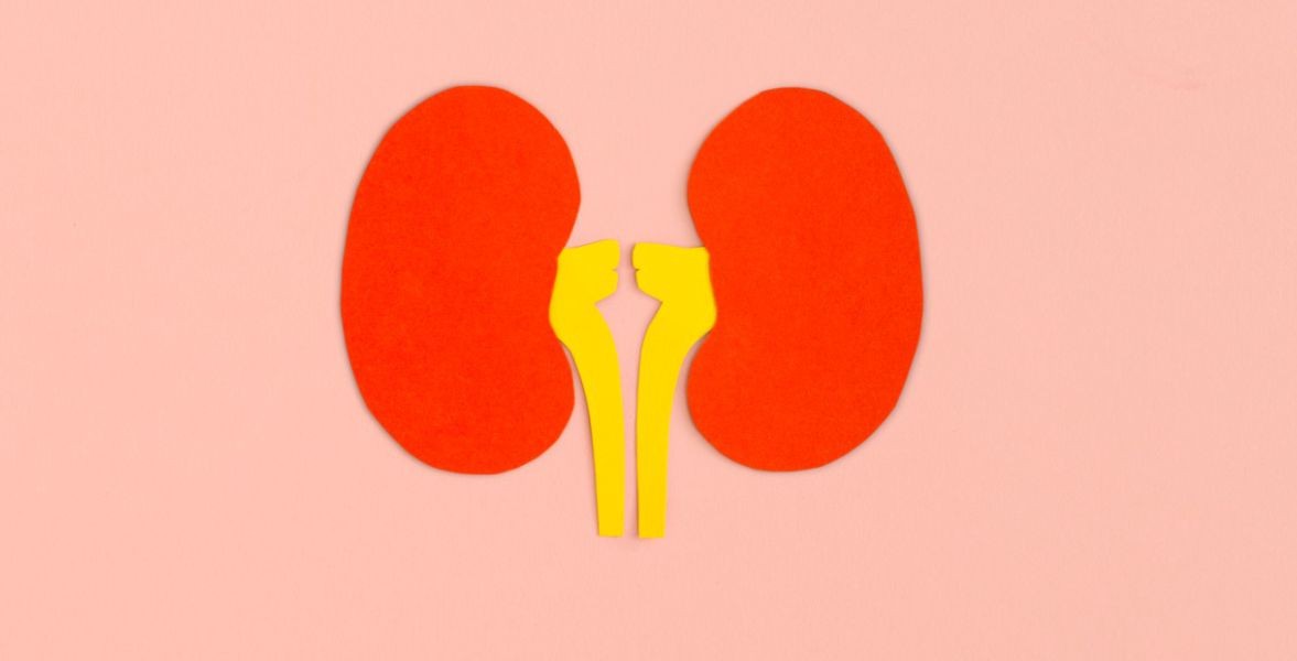
Kidney Failure – Types, Symptoms & Early Signs
Your kidneys are a pair of organs situated within the region of your lower back. The kidneys filter your blood and take away toxins from your body. These toxins move to your bladder and are eliminated via the urine. Kidneys lose their ability to efficiently filter waste from your blood in case of inflammation or damage. Many factors may interfere with the performance of the kidney and affect its health such as: · Certain acute and chronic diseases· Toxic exposure to environmental pollutants or sure medications· Severe dehydration· Insufficient blood flow to the kidneys· kidney trauma If your kidneys aren’t working properly, toxins can accumulate in the body which might be life-threatening if left untreated. Keep reading as we tend to break down everything you wish to understand regarding nephritis, - symptoms, stages, treatment, Types of Kidney Failure Acute kidney failure occurs suddenly when the kidneys suddenly stop working while chronic nephritis happens over time. The 5 types of kidney failure include: Acute prerenal kidney failure Cause - Insufficient blood flow to the kidneys. The kidneys can’t filter toxins from the blood without enough blood flow.PrognosisCan usually be cured once the cause of the reduced blood flow is determined.Acute intrinsic kidney failure -Cause - Direct trauma to the kidneys, such as physical impact or an accident. Toxin overload and ischemia due to lack of oxygen to the kidneys.Prognosis lose of the ability to function.Chronic prerenal kidney failure -CauseThere isn’t enough blood flowing to the kidneys for an extended period.Prognosis The kidneys begin to shrink and lose the ability to function.Chronic intrinsic kidney failure -Causes Long-term damage to the kidneys due to intrinsic kidney disease due to Trauma to the kidneysPrognosisEventual kidney damageChronic post-renal kidney Cause Long-term blockage of the urinary tract prevents urination.PrognosisPressure and eventual kidney damage failure.Symptoms of Kidney Failure – Early-stage kidney failure typically doesn’t cause noticeable symptoms, As per CDC, 90% of people are not even aware of their chronic kidney problem As the condition progresses, the symptoms might include: o A reduced quantity of Urineo Legs, ankles, and feet are getting swelled.o Shortness of breatho Excessive tiredness/fatigueo Persistent nauseao Confusiono Pain or pressure in your chesto Seizureso Coma Early signs of kidney failure include- o Decreased Urine outputo Swelling in limbs caused by fluid retentiono Shortness of breath
More detailsPublished - Mon, 25 Jul 2022
Search
Popular categories
Health and Wellness
231Skill Development
7Technology
5Community Impact
2Success story
2Creativity
1Latest blogs

DeepSchool: The Story of an Idea That Refused to Sit Still
Tue, 02 Dec 2025

Transforming Emergency Care: The Story Behind Rigomo's Revolutionary PPMMP Course
Sun, 12 May 2024

Empowering Rural Healthcare: How Pogiko's AI is Bridging the Gap in Medical Services
Thu, 25 Apr 2024
Write a public review