Created by - Rigomo Team
HYPONATREMIA
A condition that develops when the sodium level in the blood is too low. In this situation, the body retains an excessive amount of water. Because of this, the blood's salt levels are diluted and fall. A blood sodium level of 135 to 145 mEq/L is considered to be normal. When the blood's sodium level goes below 135 mEq/L, hyponatremia sets in.a) True hyponatremia: Serum sodium concentration < 125 mEq/L and a serum osmolality < 250 mOsm/kg.b) Pseudo-hyponatremia: Secondary to extreme hyperlipidemia, hyperproteinemia, or hyperglycemia. Although the sodium level in the body as a whole is unchanged, the serum sodium level falls.CAUSES1. Hypovolemic hyponatremiaa)Extrarenal losses: Hypovolemic hyponatremia as a result of extrarenal sodium loss, urinary sodium level < 20 mEq/L.b) Renal losses: Hypovolemic hyponatremia as a result of renal sodium loss, the urinary sodium level > 20 mEq/L.2. Euvolemic hyponatremia: Occurs as a result of an increase in total body water content; normal sodium levels are present. Euvolemic hyponatremia is brought on by hypothyroidism and the syndrome of inappropriate antidiuretic hormone.3. Hypervolemic hyponatremia: Occurs with cirrhosis, congestive heart failure, renal failure, and nephrotic syndrome.CLINICAL FEATURES The influx of water into brain cells could result in: —Apathy—Altered consciousness—Agitation—Anorexia—Coma—Headaches—Nausea—Seizures—Vomiting—WeaknessDIFFERENTIAL DIAGNOSES: Includes pseudo hyponatremia, which can be caused by diabetic ketoacidosis and a hyperosmolar state (leading to hyperglycemia), multiple myeloma (leading to hyperproteinemia), or hypertriglyceridemia (leading to hyperlipidemia)EVALUATIONa) Laboratory studies: Include a serum electrolyte panel (i.e., sodium, potassium, chloride, and bicarbonate); serum glucose, blood urea nitrogen [BUN], and creatinine levels; and urinalysis (to determine the urine sodium level and osmolality)b) Electrocardiography: An electrocardiogram (ECG) should be obtained to evaluate for arrhythmia.THERAPY1. Fluid restriction or replacementa) Hypervolemic and euvolemic hyponatremia usually result from hemodilution; When a patient is stable and asymptomatic, fluid restriction is the first course of treatment. Demeclocycline (600 to 1,200 mg/day) or furosemide can be used to treat the syndrome of inappropriate antidiuretic hormone-induced inhibition of water reabsorption.b) Hypovolemic hyponatremia can usually be treated with isotonic saline.2. Sodium replacement: Sodium deficits can be calculated as follows: Na+ deficit (mEq) = 0.6 * (weight kg) * (140 - serum Na+)a) If the hyponatremia is acute, is severe (i.e., the serum sodium level is less than 120 mEq/L), and results in CNS symptoms, administration of 3% (hypertonic) saline solution at 25 to 60 mL/hour is indicated.—The serum sodium concentration should not increase at a rate that exceeds 2 mEq/hour.—When sodium levels reach 120 mEq/L or the patient exhibits a significant clinical improvement, hypertonic saline should be stopped.b) The rate of serum sodium correction should not exceed 0.5 mEq/L/hour (12 mEq/L/day) in individuals with persistent severe hyponatremia. 3 percent saline should be given if the hyponatremia is severe (i.e., serum sodium levels are less than 120 mEq/L) and develops quickly with CNS symptoms.DISPOSITIONAdmission: Symptomatic patients, with serum sodium concentration below 125 mEq/L (with or without symptoms), patients who require intravenous or pharmacologic correction of the sodium imbalance, and patients who have significant comorbid factors (e.g., diabetes, advanced age, sepsis).Discharge: If the patient is being discharged, the case should be discussed with a primary care physician and a follow-up appointment should take place within 48 to 72 hours.
More detailsPublished - Mon, 25 Jul 2022

Created by - Rigomo Team
HYPERNATREMIA
The medical word for having too much sodium in the blood is hypernatremia. An essential ingredient for the body's healthy operation is sodium. The blood contains the majority of the sodium in the body. It is also an essential component of the lymph fluids and cells in the body. A rise in serum sodium concentration to a number above 145 mmol/L, which is considered normal between 136 and 145 milliequivalents per liter, is the definition of a common electrolyte issue. CAUSES1. Reduced water intake can be caused by a defective thirst mechanism, unconsciousness, an inability to drink, or a lack of access to water.2. Increased water loss can be caused by diarrhea, fever, diabetes insipidus, osmotic diuresis, hyperventilation, thyrotoxicosis, sweating, vomiting, and severe burns.3. Increased sodium intake or renal salt retention is due to hypertonic saline ingestion or infusion, Cushing disease, congenital adrenal hyperplasia, hyperaldosteronism and sodium bicarbonate administration.CLINICAL FEATURESSymptoms:—Lethargy/lack of energy—Increased thirst—Confusion—Muscle irritability/twitches—Seizures—Tremulousness—Weakness—ComaPhysical examination findings: —Dry mucous membranes—Flat neck veins—Orthostatic hypotension—Poor skin turgor—Respiratory paralysis —Tachycardia—Tonic spasmsEVALUATION1. Laboratory studies should include a complete blood count (CBC); serum electrolyte panel; serum glucose, BUN, and creatinine levels; urinalysis, urine sodium level, and osmolality.2. Electrocardiography: An ECG should be obtained.THERAPY1. Fluid replacement: The amount of water needed to correct hypernatremia can be estimated as follows:Water deficit (L) = 0.6* (usual body weight in kg)* (measured Na+ concentration - desired Na+ concentration) / measured Na+ concentration—To increase blood pressure and tissue perfusion in cases of severe dehydration, normal saline or lactated Ringer solution should be provided.—After the perfusion has been restored, 0.45 percent saline is given to keep the urine production at 0.5 mL/kg/hour.2. Sodium reduction: The rate of sodium reduction should not exceed 10 to 15 mEq/L/day. The goal is to reach a normal serum sodium value in 48 to 72 hours.DISPOSITIONAdmission: Symptomatic patients, with serum sodium concentration greater than 160 mEq/L (with or without symptoms), patients who require intravenous or pharmacologic correction of the sodium imbalance, and patients who have significant comorbid factors require admission.Discharge: Before discharging a patient, the case should be discussed with a primary care physician and arrangements should be made for appropriate follow-up.
More detailsPublished - Tue, 26 Jul 2022

Created by - Rigomo Team
KIDNEY FUNCTION TESTS
Healthy kidneys remove wastes and excess fluid from the blood. Blood and urine tests show how well the kidneys are doing their job in removing the toxic waste from the body.The urine tests also help figure out whether the proteins and sugar are being excreted out or not, which implies damage to the kidney itself. Here's a fast guide to the tests for kidney functionSerum CreatinineCreatinine is a waste product that is formed after muscle tissue breakdown. The levels of Creatinine in the blood will vary by age and body size. Higher levels of creatinine are indicative of kidney problemsA creatinine level of >1.2 for Females and >1.4 for Males is a sign of concern. Glomerular Filtration Rate (GFR)This is to determine, how well the kidneys work in removing wastes and excess fluid from the blood. It's calculated from the rate at which kidneys are filtering the wastes, the rate depends on age, gender, size, height, and weight. A GFR below 60 could be a sign that the kidneys aren't operating properly. Once the GFR decreases below 15, one is at high risk of needing dialysis or an organ transplant.Blood urea nitrogen (BUN)BUN quantifies nitrogen which is made from protein breakdown, in the blood. Its level is between 7 and 20. As the performance of the kidney decreases, the level rises.UrinalysisIncludes microscopic examination of a urine sample to find out the presence of molecules that are normally not present in urine such as protein, sugar, pus cell, and microorganisms Urinalysis is most commonly performed, when there is a suspicion of urinary tract infection or when the patient presents with symptoms of Abdominal pain, Back pain, Painful/frequent/ burning urination, and blood in the urine, which could point towards UTI, bladder injury or kidney disease. The test is also used to monitor diabetes, kidney or liver disease; or detecting pregnancy.A chemical examination or dipstick test is done by using strips that are infused with chemicals and dipped in urine to reveal what is present in urine and in what amount? [ low or excess]As we know lab reports specify a few tests such as · Specific gravity: it is the measure of concentration– How concentrated your urine is? A higher concentration means you are not drinking enough water..· pH: Urine is usually slightly acidic, about pH 6. Changes in pH level or acidity indicate issues o A value less than 5, that is too acidic– there is a risk of development of stoneso A value of more than 7 is basic and indicates a bacterial infection· Protein: The traces of protein in urine are normal. A small amount of increase in protein in urine can occur after exercise, fever, or stress, but a large amount of protein indicates a kidney problem.· Glucose: Glucose (sugar) is not detected normally in urine and detection is a sign of diabetes· Ketones: just like glucose, any amount of ketones detected in urine could be a sign of diabetes
More detailsPublished - Wed, 27 Jul 2022

Created by - Rigomo Team
HYPOKALEMIA
Hypokalemia describes low potassium levels in the blood. Less than 3.5 mEq/L of serum potassium is the most typical electrolyte abnormality. Your body needs potassium to function properly. The foods you eat provide potassium for your body.CAUSES1. Extra-renal causes: Inadequate dietary intake, diarrhea, vomiting, and redistribution (e.g., as a result of insulin administration, epinephrine injection)2. Renal causesa) Drug-induced renal losses: —Loop diuretics—Penicillin—Aminoglycosides—Amphotericin Bb). Hormone-induced renal losses: Can occur as a result of primary adrenal adenomas, adrenal hyperplasia, ectopic adrenocorticotropic hormone syndrome, renin-secreting tumors, renal artery stenosis, and malignant hypertensionc) Renal tubular acidosis, Bartter syndrome, or chronic magnesium depletion can also lead to hypokalemia.CLINICAL FEATURES1. Symptoms:—Paresthesias—Constipation—Weakness/fatigue—Excessive thirst—Excessive urination—Fainting /lightheadedness2. Examination findings:—Areflexia—Arrhythmias—Ileus—Orthostatic hypotension—ParalysisEVALUATION1. Laboratory studies should include a serum electrolyte panel; serum BUN, creatinine, creatinine phosphokinase, phosphate, magnesium, and glucose levels; and urinalysis.a) Creatinine phosphokinase levels in the serum may be high.b) Urinalysis: The urine specimen may be dipstick-positive for red blood cells. Myoglobin may be seen in a formal urinalysis, which is consistent with rhabdomyolysis.2. Electrocardiography: ECG findings include T-wave flattening or inversion, U waves, ST-segment depression, premature ventricular contractions, and a wide QRS complex.THERAPY1. Potassium replacementa) If the serum potassium level is higher than 2.5 mEq/L and there are no abnormalities on the electrocardiogram (ECG), 40 to 80 mEq of potassium chloride should be given every day until the imbalance is resolved, with no more than 40 mEq given in a single dosage.b) For severe hypokalemia, defined as a potassium level of less than 2.5 mEq/L, 10 mEq of potassium chloride are infused hourly through intravenous piggyback in 50–100 mL of 5% dextrose in water or normal saline for 3–4 hours.—No more than 40 mEq of potassium should ever be put in a single liter of intravenous fluid, and no more than 10 mEq should be given per hour.—Continuous cardiac monitoring is required.2.Magnesium replacement with 2 g of magnesium sulfate in 50 mL of 5% dextrose in water administered over 20 minutes may be necessary.3.Phosphate replacement: Potassium phosphate may be used instead of potassium chloride if the serum phosphate level is low. The 2.5 mg/kg daily dosage is advised.DISPOSITIONAdmission—Patients with serum potassium concentrations of less than 2.5 mEq/L require admission to the hospital.—Patients with malignant cardiac dysrhythmias, digitalis toxicity, profound weakness with impending respiratory failure, rhabdomyolysis, hepatic encephalopathy, or a serum potassium level of less than 2.0 mEq/L require admission to the intensive care unit.Discharge: Patients with mild hypokalemia (serum potassium concentration = 2.5 to 3.5 mEq/L) can usually be managed as outpatients with gradual oral potassium repletion, provided they do not have ECG abnormalities, or profound muscular weakness, ileus, or other serious effects. Patients who are discharged on oral supplementation should have a follow-up appointment within 48 to 72 hours.
More detailsPublished - Thu, 28 Jul 2022

Created by - Rigomo Team
HYPERKALEMIA
The medical term for an elevated potassium level in your blood is hyperkalemia. A molecule called potassium is essential for the health of your heart's muscle and nerve cells. Normal blood potassium levels range from 3.6 to 5.2 millimoles per litre (mmol/L), while hyperkalemia is caused by serum potassium levels that are higher than 5.5 mEq/L.CAUSES1.Extra-renal causes: Insulin deficiency, acidemia, hyperosmolality, Beta blocker administration, oral or intravenous potassium supplements, penicillin potassium salts, massive blood transfusion, crush injuries, burns, mesenteric or muscular infarction, and tumor lysis syndrome.2.Renal causes: Chronic renal insufficiency, acute renal failure, hypoaldosteronism, and drugs (e.g., nonsteroidal anti-inflammatory drugs, cyclosporine, heparin, angiotensin-converting enzyme inhibitors, potassium-sparing diuretics) can also cause hyperkalemia.3.Hyperkalemia is most frequently caused by laboratory error.CLINICAL FEATURESSymptoms:—Confusion —Paresthesias/Muscle weakness /numbness in —limbs—Weakness—Abdominal pain and diarrhea.—Chest pain.—Irregular, fast/fluttering heartbeat—Nausea/vomiting.Physical examination findings: —Areflexia—Cardiac arrest—Ileus—Paralysis—Respiratory insufficiencyDIFFERENTIAL DIAGNOSES include pseudo-hyperkalemia, which can occur as a result of hemolysis, extreme leukocytosis, acidosis, thrombocytosis, or cold agglutinins.EVALUATION1.Laboratory studies include a serum electrolyte panel, serum BUN, creatinine, glucose, and magnesium levels.2.Electrocardiography: —An ECG should be taken and the patient should be put on a continuous heart monitor.—Peaked T waves and a shorter QT interval are early ECG abnormalities.—Later, enlarged QRS complexes, protracted PR intervals, small P waves, and elevation or depression of the ST segment are observed. —In addition to a sine wave pattern, ventricular fibrillation, and asystole, advanced modifications include absent P waves, distinct QRS complex enlargement, and tall T waves.THERAPY1.Acute therapy:a)Calcium chloride or calcium gluconate is administered over 2 minutes and repeated in 5 to 10 minutes if necessary. Calcium is the most crucial first step in treatment because it prevents malignant arrhythmia by stabilising cell membranes without affecting potassium levels. Calcium chloride should ideally be administered through a central line since if it accidentally leaks from peripheral intravenous lines, it severely damages skin.b) Sodium bicarbonate (44 mEq [one ampule] administered intravenously over 5 minutes and repeated 10 to 15 minutes later if necessary) causes an intracellular influx of potassium. Onset of action occurs in approximately 15 minutes.c) Regular insulin (10 to 20 U administered via an intravenous push) with 10% dextrose (500 mL in water) administered over 1 hour, or 10U of insulin administered via an intravenous push with one or two ampules (25 g) of 50% glucose administered over 5 minutes will lower potassium by causing an intracellular shift. Effects occur 30 to 60 minutes after administration.d)Furosemide, bumetanide, and acetazolamide all increase potassium excretion.e) Dialysis should be considered for patients with severe hyperkalemia who have failed to respond to pharmacologic attempts at lowering the potassium level and for patients with acute or chronic renal failure.2. Maintenance of potassium balance. Potassium balance is maintained by:a) Diuretics and fludrocortisoneb) Cation-exchange resins, such as sodium polystyrene sulfonatec) Aldosterone, either as desoxycorticosterone acetate or fludrocortisone acetate.DISPOSITIONAdmission: When ECG abnormalities or clinical manifestations of hyperkalemia are present, admission to the intensive care unit with continuous cardiac monitoring is required.Discharge: Patients with mild serum potassium elevations in the absence of clinical and ECG abnormalities can be discharged, provided any identifiable predisposing factors have been corrected. Patients should have a follow-up evaluation within 48 to 72 hours.
More detailsPublished - Thu, 28 Jul 2022

Created by - Rigomo Team
How to manage patients in Unconsciousness state
The state of Unconsciousness occurs when a person is not able to respond to people and activities Somebody, who is unconscious cannot cough, clear his/her throat, or flip his/her head if in distress. Once unconscious, someone is in peril of choking, creating it vital to stay the airway clear while awaiting medical aid.If you'll be able to verify what caused the loss of consciousness, call emergency medical services and provide care for that cause/injury till they arrive. If you can not verify what has caused the loss of consciousness, offer care for general first aid care and call emergency medical services if the person doesn't revive promptly (i.e., at intervals of a few minutes).Check the person's airway, breathing, and circulation.If you are not suspecting a spinal injury, place the person in the recovery position:Turn the person's face toward you.Kneel beside the person Make sure that both of the person’s legs are straight.Bring the arm that is farthest from you across the person’s chest, and hold the back of her hand against the cheek nearest to you.Keeping the person’s hand pressed against her cheek, pull on the far leg and roll the person towards you and onto her side.Adjust the upper leg so that both the hip and the knee are bent at right angles.If necessary, adjust the hand under the cheek to make sure that the head remains tilted and the airway stays open. After 30 minutes turn the person to the other side.Keep the person heated till emergency medical facilities arrive.
More detailsPublished - Thu, 28 Jul 2022

Created by - Rigomo Team
Types of poison and their adverse effect
If the person has no symptoms even though he/she may have consumed the poison, then it is important to immediately reach the hospital or emergency room for poison treatment Poison is something that kills or injures the cells/tissues through its chemical actions. Most poisons are ingested. The word poison comes from the Latin word – “potare” - assuming to drink. However, poisons can even enter the body in other ways: Through inhalation [spreads via respiratory] Through the skin By IV injection From exposure to radiation Venom from a snake bite Poisons embrace contact with extremely toxic chemicals not meant for human intake like cyanide, paint thinners, or unit cleansing merchandise.Many substances that are meant for human consumption such as a few foods and medicines, can be lethal if consumed in excess Foods· Some mushrooms are toxic· Drinking contaminated water· Food that has not been properly handled· Drugs· Drugs that are useful in therapeutic doses could also be deadly if taken in excess. Examples include:Drugs Beta-blockers: Beta blockers are used to treat heart conditions (for example, angina, and abnormal heart rhythms) and other conditions, (for example, high pressure, sick headache bar, phobic disorders) In excess, they can cause problems. Warfarin (Coumadin): Anticoagulant is the active ingredient in several rat poisons and could cause serious injury and death if an excessive amount is taken. Vitamins: Vitamins, particularly A and D, if taken in large amounts can cause toxicity. Signs and symptoms of poisoning are variable and there is no simple way to classify them. · Enlarged/dilated pupils · Excessive drooling/Dry mouth and skin. · Some increase the respiratory rate, whereas others may slow it. · Some are painless and few are painful · Some may cause loss of consciousness
More detailsPublished - Fri, 29 Jul 2022

Created by - Rigomo Team
WHAT IS THE EMERGENCY MANAGEMENT FOR HYPOGLYCEMIA
Your blood sugar (glucose) level is below the normal range (less than 50 mg/dL) if you have hypoglycemia. Glucose serves as your body's main energy source. Diabetes control and hypoglycemia commonly go hand in hand. However, people without diabetes can experience low blood sugar as a result of a wide range of illnesses and treatments, many of which are unusual.Etiology: Insulin (secreted by the pancreas), counter-regulatory hormones (such as glucagon, catecholamine, and glucocorticoids), and growth hormones interact intricately to maintain glucose homeostasis. Hypoglycemia is often categorized as postprandial (reactive) or fasting.1. Spontaneous hypoglycemia:a) Postprandial (reactive) hypoglycemia is characterized by declining glucose levels after a glucose load.b) Alimentary hypoglycemia occurs in patients who have recently undergone gastrointestinal surgery.c) Pre-diabetic glucose intolerance: Hypoglycemia may be an early manifestation of non–insulin-dependent diabetes mellitus.d) Functional (idiopathic) hypoglycemia: Hypoglycemia occurs between the fed and fasting state.e) Fasting hypoglycemia occurs in patients with significant underlying pathologic conditions.f) Endogenous insulin excess may result from insulinomas (nonmalignant pancreatic tumors) or extrapancreatic neoplasms.g) Regulatory hormone deficiencies: Acquired or congenital deficiencies of glucagon, glucocorticoids, or growth hormone can lead to hypoglycemia.h) Organ failure: Impaired liver or kidney function can lead to hypoglycemia.Systemic disease: Shock, sepsis, and starvation can cause hypoglycemia.2. Induced hypoglycemiaa) Insulin-induced hypoglycemia, seen in patients with diabetes, is the most common cause of hypoglycemia seen in the emergency department.b) Factitious hypoglycemia may be seen in psychiatric patients (e.g., as a result of the ingestion of oral hypoglycemic agents by a patient with Munchausen syndrome).c) Chemical-induced: Alcohol-induced hypoglycemia is found in malnourished, alcoholic patients. Hypoglycemia can also be brought on by other substances and medicines.RISK FACTORS: Young children and elderly patients are at high risk for the development of hypoglycemia.CLINICAL FEATURES are caused by the direct effects of hypoglycemia on the central nervous system as well as its indirect effects on the sympathetic nervous system. Patients may be asymptomatic, or they may present with a wide variety of symptoms.1. Nonspecific systemic symptoms include dyspnea, hypertension, hyperventilation, sweating, palpitations, peripheral vasodilation, pallor, tachycardia, and tremulousness.2. Neurologic symptoms include paresthesia, neurologic deficit, diplopia, clonus, and transient hemiplegia.3.Psychiatric manifestations include impairment of memory, change of personality, combative behavior, fatigue, headache, insomnia, nightmares, visual problems, catatonia, convulsions, and general sluggishness.DIFFERENTIAL DIAGNOSES: Hypoglycemia may masquerade as a neurologic, psychiatric, or cardiovascular disease. Stroke, diabetic ketoacidosis, nonketotic hyperosmolar coma, alcohol intoxication, alcohol withdrawal, and other causes of coma must all be ruled out.EVALUATION: Testing for glucose at the bedside is a good method of excluding hypoglycemia.THERAPY1. Prehospital providers commonly administer one ampule (25 g) of 50% dextrose intravenously. Alcoholic patients should receive thiamine (100 mg administered intravenously) before receiving dextrose to prevent Wernicke-Korsakoff syndrome. If intravenous access is unavailable, glucagon (0.5 to 2.0 mg) is administered intramuscularly or subcutaneously. The glucagon dose can be repeated twice.2. After initial treatment, a meal rich in complex carbohydrates should be given. A continuous intravenous infusion of water containing 5% dextrose should be started if the patient is unable to swallow.DISPOSITIONAdmission is indicated for patients who have taken an oral hypoglycemic agent or long-acting insulin. Patients in whom no obvious cause for the hypoglycemia can be identified and patients with persistent neurologic deficits or cardiac complications (e.g., coronary or cerebrovascular insufficiency) should also be admitted.Discharge: Patients whose symptoms are rapidly reversed without complications and in whom a clear cause for the hypoglycemia has been identified may go home. Patients with diabetes who experience a hypoglycemic episode should be taught how to adjust their insulin dose, food intake, or both based on their level of physical activity.
More detailsPublished - Fri, 29 Jul 2022

Created by - Rigomo Team
Dos and Don’ts during Dog Bite
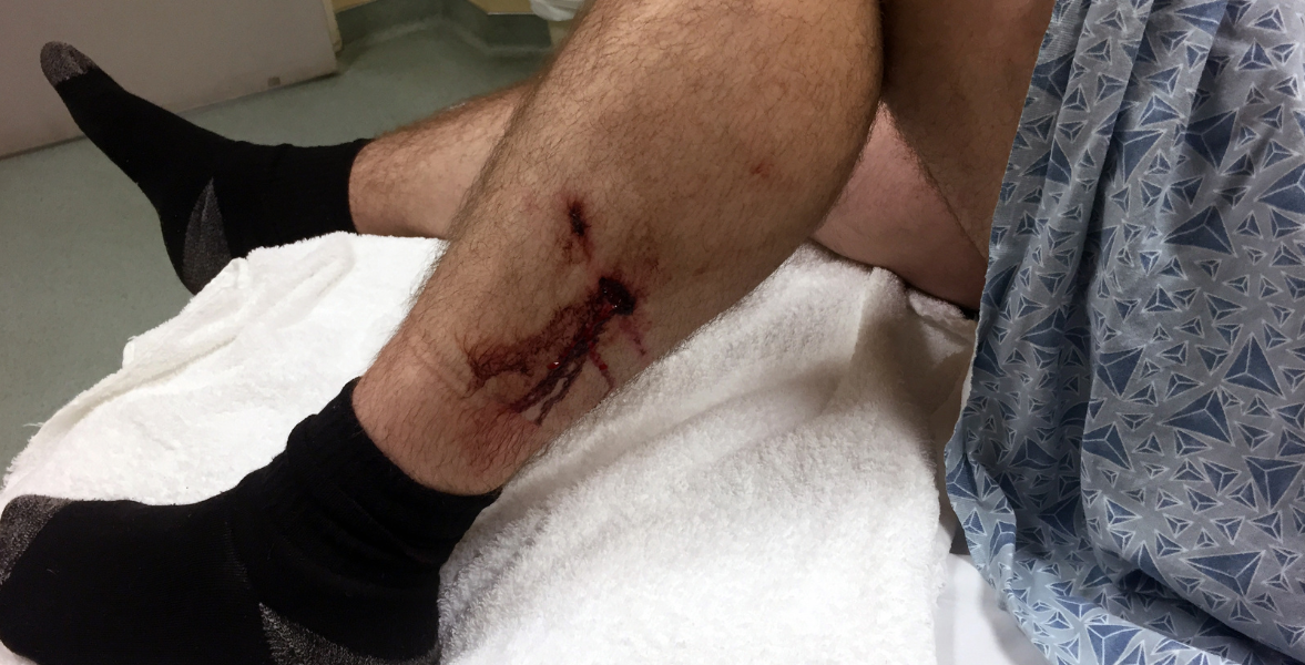
Dog bites account for up to 90% of all animal bites. 4.5 million dog bites occur every year within the U.S., and quite 27,000 victims need surgical intervention. Injuries could be deep that involve structures such as skin as well as muscles, bones, nerves, and blood vessels. It could result in dangers of acquiring infections, or even tetanus after a bite.Wound cleansing decreases the danger of infections from dog bites. After confirming that the victim is medically stable, physicians begin a primary assessment by taking a history. Many medical conditions place a patient at high risk of wound infection from a bite Dogs have rounded teeth, and it's the pressure exerted by their jaws may cause vital injury to the tissues beneath the skin, as well as bones, muscles, tendons, blood vessels, and nerves. The risk of being bitten by a dog will increase if there's a dog within the home. Males are more frequently bitten by dogs than girls. Children between the ages of five and nine are bitten by a dog more than adults and they need medical attention. Dos during Dog Bite -If a dog bites you or somebody with you, take the person to a secure place far from the dog to stop any further attack and injury. Infants and kids should be evaluated for any bite. If there aren't any signs of skin injury or if there's any abrasion from a bite, it will be great to observe for signs and symptoms of infection (pain, redness, warmth, swelling, and emptying of pus or fluid) before seeking treatment.If the skin is unbroken, clean the bite with water.Gather data from the dog's owner concerning the dog's immunization status if possible.Since dog bites can cause vital injury to structures under the skin, a kind of injury that can't invariably simply be seen. Moreover, if there's pain at or close to the bite, underlying tissues and alternative structures are affected, or if the bite disrupts the skin by inflicting a puncture, laceration, or tear, immediately visit a doctor or Emergency Department. Prevention – Dog bites usually occur once there's miscommunication between the dog and the victim. It’s not common to have an unmotivated attack by a stray dog. Often, it's the dog owner or a loved one who is bitten. To stop being bitten by a dog: Choose a dog breed that's compatible with the family. Aggressive dogs might not be suitable in the home with infants and little youngsters. Dogs are social animals; thus, socialization and coaching can facilitate and minimize the danger of dog bites. Do not approach a stray or strange dog. Do not approach a dog with fast motions/speed. Give some time for the dog to acknowledge your presence before attempting to pet it.Will I get rabies?If the dog is not immunized, take an antirabies injection right after the bite along with a Tetanus shot. If treatment is initiated promptly following a bite, rabies can be prevented. If exposure isn't treated and a person develops clinical signs, he/she may sometimes end up in death.
More detailsPublished - Sat, 30 Jul 2022
Search
Popular categories
Health and Wellness
231Skill Development
7Technology
5Community Impact
2Success story
2Creativity
1Latest blogs

DeepSchool: The Story of an Idea That Refused to Sit Still
Tue, 02 Dec 2025

Transforming Emergency Care: The Story Behind Rigomo's Revolutionary PPMMP Course
Sun, 12 May 2024

Empowering Rural Healthcare: How Pogiko's AI is Bridging the Gap in Medical Services
Thu, 25 Apr 2024
Write a public review