Created by - Rigomo Team
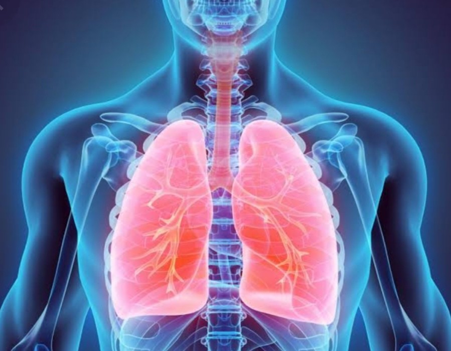
Non-cardiogenic Pulmonary Edema (NCPE)
Non-cardiogenic pulmonary edema (NCPE) is also known as acute respiratory distress syndrome. NCPE is a form of pulmonary edema resulting from an abnormal increase in the permeability of pulmonary vascular membranes.CAUSES of NCPE include sepsis, aspiration of gastric contents, near-drowning, thermal injury, trauma, high-altitude pulmonary edema, radiation, transfusions, eclampsia–preeclampsia, and selected drug reactions or overdoses, including narcotic and aspirin overdose.PATHOGENESIS: Cardiovascular, hydrostatic, or hemodynamic edema is unrelated to NCPE. The proposed mechanism for the edema is damage to the vascular endothelium as a result of complement cascade activation or direct damage by bacterial endotoxin. Pulmonary surfactant is disrupted and lung compliance is decreased.CLINICAL FEATURES1. Symptoms: Tachypnea that progresses quickly with dyspnea is common.2. Physical examination findings include bilateral rales.DIFFERENTIAL DIAGNOSES Most frequently, NCPE is confused with pneumonia or CHF. Asthma, COPD, pulmonary embolism, an anaphylactic reaction, and foreign body aspiration should also be considered.EVALUATION1. Chest radiographs, ABGs, and an ECG should be obtained for all patients suspected of having NCPE.a) The chest radiograph shows a normal-sized heart and bilateral patchy alveolar infiltrates. In extreme situations, the infiltrates could cause the lungs to "white out.”b) The ABG demonstrates hypoxia and an increased alveolar-arterial gradient where PaO2/FIO2 is less than 300.2. Laboratory tests: Pertinent laboratory tests include a complete blood count (CBC), urinalysis, an electrolyte panel, and blood urea nitrogen (BUN) and creatinine levels.THERAPY1. To keep the oxygen saturation level above 90%, more oxygen should be given. In intubated patients, the FIO2 should be kept below 50%, if possible, because of the potential for pulmonary oxygen toxicity.2. Intubation and mechanical ventilation are indicated for patients who, despite supplemental oxygen, have inadequate oxygenation or ventilation. PEEP should be added if adequate oxygenation cannot be maintained with an FIO2 of less than 50% to keep tidal volumes low at < 6 mL/kg.3. Treatment of the underlying disorder should be undertaken (e.g., antibiotics for sepsis, alkalinization, and dialysis for aspirin overdose).4. The use of corticosteroids, non-steroidal anti-inflammatory drugs, and anticoagulants is being investigated for the future treatment of acute respiratory distress syndrome.DISPOSITION1. Admission: The vast majority of patients with NCPE will need to be admitted to the hospital and should be sent to a monitored bed.a) Many patients who develop NCPE do so as a result of medical conditions that often require hospitalization in and of themselves (e.g., sepsis, near drowning, thermal injury, trauma, aspirin overdose).b) Patients' respiratory conditions may quickly deteriorate and NCPE may worsen quickly. Patients who are intubated or seriously unwell must be admitted to the ICU.2. Discharge: Overdose patients who are not hypoxic and are asymptomatic after 6 to 12 hours of observation are usually discharged home.
More detailsPublished - Thu, 06 Oct 2022

Created by - Rigomo Team
Physical fitness – An eminent component of Overall Well-being
Physical fitness is defined as “one’s ability to maintain daily activities with fast performance, endurance, and proper strength as well as fewer chances of falling ill, or having fatigue, and stress which could lead to inactive behavior.” This also embodies flexibility and the ability to run quickly or lift weights with ease. This article provides details of the five main components of fitness.Fast facts on fitness:o Maintaining physical fitness can facilitate forestalling some conditions.o With exercise, body mass index can be modified o Athletes’ hearts rate differ as per their chosen sport.o Muscle strength can increase due to fiber hypertrophyo Stretching to increase flexibility can ease lots of medical complaints related to the stiffness.Flexibility is important as it improves the ability to perform movements smoothly and would possibly facilitate forestall injuries. Tightness of ligaments and tendons around the joint due to inactivity can cause stiffness which could lead to pain and difficulty while moving the joint. Many activities can elongate and stretch the ligaments, and tendons thereby increasing flexibility.There are three common sorts of stretches that folk use to increase flexibility:o Dynamic stretching: This refers to the ability to perform the full range of motion. People use this type of stretch as a warm-up exercise as it helps prepare the body for physical activity.o Static-active stretching: This refers to holding the body in a much-stretched position and maintaining that position for some time. One example of static-active stretching is the splits.o Trajectory stretching: People got to entirely move into a stretching exercise, once the body is already warmed-up and limber from exercise. It involves stretching in various positions. The stretching boosts flexibility. Having a daily stretching program is usually the most effective way of achieving whole-body flexibility and better quality of life.
More detailsPublished - Thu, 06 Oct 2022
Created by - Rigomo Team
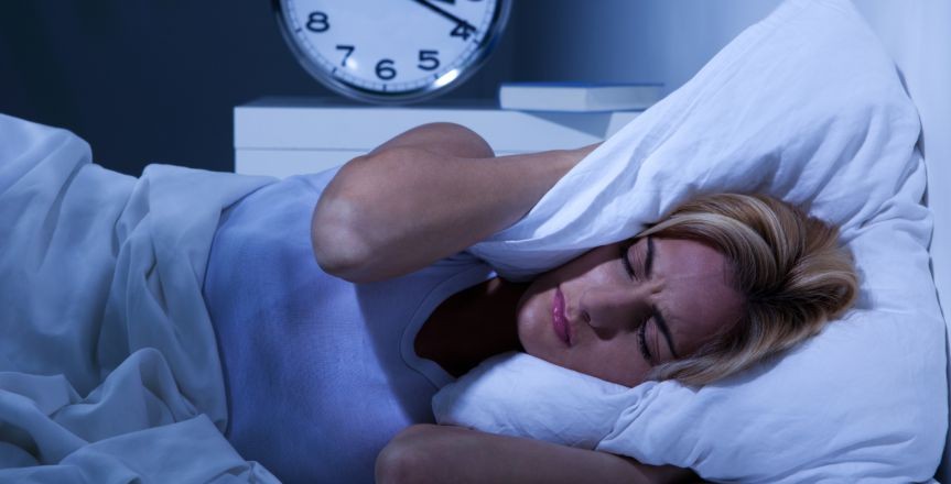
Acupressure for insomia
Acupressure has recently come back to the fore as another remedy for the sleep disorder. Instead of merely inducing sleep, it conjointly attempts to treat a number of the associated causes of sleep disorder, like anxiety, and stressHere’s a comprehensive guide to exploiting G-Jo [The technique that combines massage and acupuncture] to attain deep and quality sleep.Insomnia is defined as an inability to fall asleep or to remain asleep. sleep disorder sufferers typically have symptoms like fatigue and low energy, poor mood, and difficulty concentrating, which may affect performance at work.Acupressure is a kind of medical practice that has been used for thousands of years, especially in China. It is a combination of “acupuncture” and “pressure,” during which specific points (“acupoints”) on the body are aroused manually by applying pressure in a particular way. The pressure is imparted through fingers, palms, elbows, and feet, or with specialized G-Jo tools.It supports the thought of energy flowing via particular routes (“meridians”) through the body. It’s believed that these meridians conjointly form a network between different organs of the body. G-Jo aims to rectify disorders by clearing blockages of energy of these meridians, and is used for ages to treat chronic muscle pain, headaches, nausea, and sleep disorders.How Will It Work to Cure Insomnia?G-Jo aims to permit higher energy flow (qi) around the body and to manage and balance the opposing forces of positive (yang) and negative (yin) energy. It is dedicated to renewing energy flows and balances where imbalance and problems with flows could have resulted in various ailments. Insomnia could happen when there is no associated physical complaints/disorder typically so treating it will be complicated. G-Jo claims that Sleep disorders with no apparent physical cause could be due to religious and emotional factors and can be effectively treated through G-Jo, as compared to other prescribed medications.o B38. The B38 or important Diaphragm acupoint is found at the level of the heart, however in the back, between shoulder blades. It helps balance emotions, that is right if sleep is being hindered by anxiety, guilt, stress, or depression. You can self-stimulate this point by using lawn tennis balls while lying on the ground.o The Pericardium 6, conjointly known as the Neiguan, is found on the inner wrist about an inch from the wrist joint crease. It certainly claims to treat each anxiety and digestion issue – each of which may contribute to a sleep disorder or poor sleep. Some practitioners say it will provide pretty much immediate relaxation.o The Heart 7 purpose, also known as the Shenmen, is found at the wrist joint crease below the small finger. It’s alleged to calm and help with a sleep disorder that is caused by over-excitement or an excessive amount of energy.o The Bladder 10 is found half an inch below the base of the skull and at the top of your neck. It’s claimed to assist relieve stress and exhaustion, which is caused by overtiredness that contributes to a sleep disorder.o The Shimein point, at the center back of the heel, can be aroused with a little item [sort of a pen] for generalized sleep disorder relief. Be cautious about performing it because if it is not done properly, it could cause hurt. You can seek advice from a trained professional on its stimulation before trying. o The Anmian point is found slightly below the ear, where the neck and jawline connect. Press the point for fifteen to twenty minutes with the index and middle finger, to induce deep and quality sleep
More detailsPublished - Sat, 08 Oct 2022
Created by - Rigomo Team
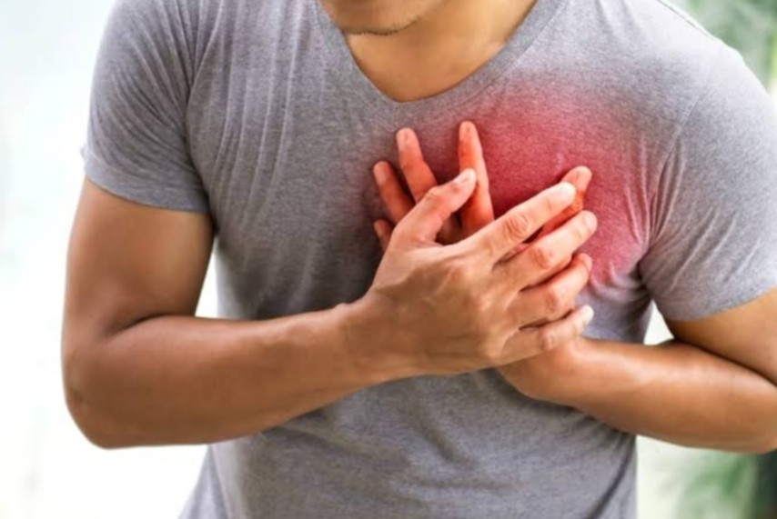
Cardiogenic Shock: Causes, Signs & Treatment
Cardiogenic shock occurs when the heart is unable to maintain perfusion adequate for the metabolic demands of the tissues.CAUSESThe cause of cardiogenic shock is usually acute MI, especially after extensive infarction of the anterior ventricular wall. In patients with smaller infarcts, complications of acute MI, such as papillary muscle rupture, septal rupture, or right ventricular infarct, can cause cardiogenic shock. Myocarditis, cardiomyopathy, and valvular stenosis are further reasons.CLINICAL FEATURES1. History: Most patients are unable to give a history, but efforts should be made to gather information from fire-rescue personnel, family members, or other witnesses.2. Physical examination findings include hypotension and tachycardia. Normal symptoms include diaphoresis, chilly extremities, and inadequate capillary refill. Breath sounds may be clear initially, or rales from acute pulmonary edema may be present. An S3 gallop or a murmur from a ruptured papillary muscle, acute mitral regurgitation, or septal rupture may be heard.DIFFERENTIAL DIAGNOSES include other causes of shock, cardiac tamponade, primary CHF, adult respiratory distress syndrome, asthma, and pulmonary embolism. A cardiac etiology is suggested by a patient in shock with risk factors for ischemic heart disease and signs of acute CHF.EVALUATION1. Electrocardiography: ST-segment elevation may be observed. Right-sided leads may show a right ventricular infarct pattern, which mandates therapy that differs from therapy for other causes of cardiogenic shock.2. Radiography: A chest radiograph may appear normal initially or show signs of acute CHF.3. Bedside echocardiography is useful for demonstrating poor left ventricular function, assessing valvular integrity, and ruling out other causes of shock, such as cardiac tamponade.4. Laboratory studies: Blood studies are not of use in making the initial diagnosis but must be included in the overall evaluation. Cardiac enzyme studies, a CBC and serum electrolyte panel, BUN and creatinine levels, and coagulation studies should be ordered. The level of serum B-type natriuretic peptide is an indicator of CHF and is a prognostic indicator of survival.THERAPY1. Emergent therapy is aimed at hemodynamically stabilizing the patient with oxygen, airway control, and intravenous access. An effort should be made to maximize left ventricular function.2. Volume expansion: If there is no sign of volume overload or pulmonary edema, volume expansion with 100-mL boluses of normal saline every 3 minutes should be tried until either adequate perfusion is restored or pulmonary congestion occurs. Right ventricular infarct patients require markedly higher filling pressures to maintain appropriate cardiac output.3. Inotropic supporta) Patients with mild hypotension (i.e., a systolic blood pressure of 80 to 90 mm Hg) and pulmonary congestion are best treated with dobutamine (2.5 µg/kg/min, titrating upward by 2 to 3 µg/kg/min at 10-minute intervals). Dobutamine supports inotropy while very slightly raising the myocardial oxygen demand.b) Patients with severe hypotension (i.e., a systolic blood pressure less than 75 to 80 mm Hg) should be treated with dopamine.— The effects of this medication depend on the dose. At doses of 2.5 to 10 µg/kg/min, it has positive inotropic and chronotropic effects. At dosages greater than 5.0 µg/kg/min, α-adrenergic stimulation gradually increases, causing peripheral vasoconstriction. At doses greater than 20µg/kg/min, dopamine increases ventricular irritability without additional benefit. Not all patients have the characteristic dose-related effects of dopamine.— A combination of dopamine and dobutamine is an effective therapeutic strategy for cardiogenic shock, minimizing the unwanted side effects of dopamine at high doses and providing inotropic support.c) If additional support for blood pressure is needed, norepinephrine, which has much stronger α-adrenergic effects, can be tried. The initial dose ranges from 0.5 to 1µg/min.d) Mechanical support (e.g., aortic counterpulsation) may be an option while arranging for more definitive management strategies.4. Reperfusion therapy: Patients with acute MI and cardiogenic shock can only be treated effectively by reperfusion of the ischemic myocardium. The recommended modality is emergent PTCA.DISPOSITIONFacilities without angiographic support should consider transferring the patient to a facility with a cardiac catheterization laboratory and cardiac surgery services.
More detailsPublished - Sat, 08 Oct 2022
Created by - Rigomo Team
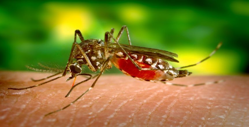
All you want to know about Dengue Fever
Dengue fever is an illness caused by the bite of a female mosquito (Aedes aegypti) carrying one among four forms of dengue fever virus (DENV). The virus is most typically found in tropical and semitropic regions, as well as Central and South America, Africa, parts of Asia, and also the Pacific Islands.Dengue isn’t contagious from person to person except it can pass from a pregnant person to a kid. Symptoms are usually mild with the 1st infection, however, if you get another infection with DENV, your risk of severe complications goes up.Can you be Immune against dengue fever?Yes, you'll get immunity to a version of the dengue fever virus once you’ve been infected with it. But you are not immune to other viral strains .. so to get full protection one must get infected with all strains to get full protection, but it is very complicated. Antibodies are formed when an infection occurs as an immune response, once your body is aware of a way to fight that specific virus, you're unlikely to get severe sickness from that virus.After obtaining one among the four strains of DENV, you don’t get ready to have dengue again, once more, especially with different strains as this new strain may get caught by antibodies generated by a different strain initially. Now, the viruses are not destroyed, but pulled inside the cells and may cause more harm leading to serious illness.What are the symptoms of dengue fever?Most dengue fever infections don’t cause symptoms. If you have symptoms, high fever (104°F/40°C) is typical, along with:o Rash.o Intense pain behind your eyes.o Nausea or acid reflex.o Muscle, bone, and joint pain.Dengue fever symptoms classically appear 4-10 days after a mosquito bite. The symptoms may last for 3 to 7 days. It has been observed that 1 in 20 individuals with dengue fever can develop severe dengue symptoms.How do I manage the symptoms of dengue fever?Dengue is managed by providing symptomatic treatment under the guidance of a healthcare provider which may include: • Keeping yourself hydrated by drinking lots of water and fluids.• Taking enough rest.• Treat pain with acetaminophen • Do not take Ibuprofen or aspirin as it might increase the risk of internal bleeding
More detailsPublished - Mon, 10 Oct 2022
Created by - Rigomo Team
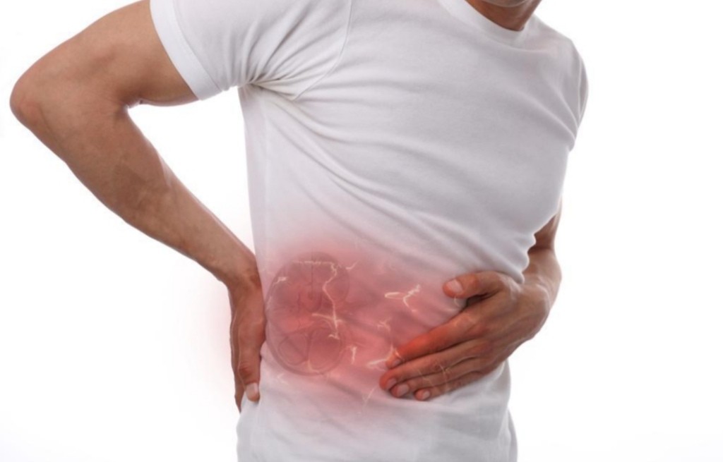
Nephrolithiasis: What is it, Types, Clinical features and Treatment
Nephrolithiasis, or kidney stones, are crystal concretions that typically develop in the kidney and are also known as renal calculi. Calculi should ideally develop in the kidneys and painlessly exit the body through the urethra. Larger stones are, however, uncomfortable and may not pass on their own, and can require surgery.Types of Renal Calculi (Stones)— Calcium oxalate and calcium phosphate stones account for 70% to 80% of all renal calculi.— Struvite (magnesium–ammonium phosphate) stones account for 10% to 15% of all renal calculi.— Uric acid stones account for 5% to 10% of all renal calculi.— Xanthine and cystine stones make up a minor percentage of renal calculi.1. Frequent sites of calculus obstructiona) Ureteropelvic junctionb) Pelvic brim: The ureter crosses the iliac vessels and narrows to 4 mm in diameter at the pelvic brim.c) Ureterovesical junction: The most common site of obstruction.d) Renal pelvis: Struvite stones commonly form large staghorn calculi, which are too large to enter the ureter and, instead, fill the renal pelvis and form the large staghorn calculus.2. Predisposing factorsa) Certain clinical conditions may predispose to stone formation (e.g., inflammatory bowel disease, immobilization, gout, hyperparathyroidism). Struvite stones are associated with chronic infection of the urinary tract by urea-splitting bacteria (e.g., Proteus, Pseudomonas, Klebsiella).b) Dietary conditions: Excess dietary oxalate, vitamin C abuse, or excess dietary purine can predispose the patient to the development of renal calculi.c) Medications— Acetazolamide, antacids, and ascorbic acid may predispose to the development of calcium stones.— Treatment with hydrochlorothiazide may cause the formation of uric acid stones.— The development of xanthine stones has been linked to allopurinol medication.CLINICAL FEATURES1. Symptomsa) Pain: The sudden onset of acute, severe, colicky pain is most commonly observed. The pain is located in the flank or abdominal area and radiates to the corresponding testicle or labia. The patient is in obvious pain and is often moving around the bed trying to find a comfortable position.b) Common symptoms include diaphoresis, nausea, and vomiting.2. Physical examination findingsa) In addition to discomfort, tachycardia, tachypnea, and hypertension are frequently seen.b) Costovertebral angle tenderness and mild abdominal tenderness without peritoneal findings are often present.c) Fever should raise suspicion of infection.DIFFERENTIAL DIAGNOSES include appendicitis, cholecystitis, diverticulitis, salpingitis, pelvic inflammatory disease, tubal-ovarian abscess, ovarian cyst or torsion, ectopic pregnancy, abdominal aortic aneurysm, and pyelonephritis.EVALUATION1. Laboratory studiesa) Urinalysis: Hematuria is usually present but not always.b) CBC: A CBC could show leukocytosis brought on by discomfort or infection.c) Basic metabolic panel: Keeping a tight eye on creatinine.2. Computed tomography (CT) 3. Radiographs: A kidney-ureter-bladder (KUB) X-ray can be obtained in some patients instead of CT. Much less sensitive and specific than CT, however drastically less radiation exposure to the patient. Generally should consider in patients with a known history of uncomplicated kidney stones.4. Ultrasound: This may be useful in pregnant patients, children, or patients with a known history of uncomplicated stones. Ultrasound is becoming more of the first-line test for hydronephrosis and/or calculus due to its superior safety profile for patients.THERAPY1. Parenteral analgesia with narcotics and a non-steroidal anti-inflammatory drug is often required.2. Antibiotic therapy: If an infection is present, justified.3. Intravenous hydration with an isotonic fluid (e.g., normal saline or lactated Ringer solution) may be beneficial.4. Ondansetron and other antiemetics are frequently required.5. Tamsulosin and other alpha-blocking medications can speed up stone movement.DISPOSITION1. Admissiona) Patients with uncontrolled pain, persistent emesis, or concomitant infection should be admitted to the hospital.b) Patients with 1 kidney only should be admitted.c) Patients with big or near-big stones might need to be admitted to the hospital.i) Stone size— 90% pass if less than 4 mm— 50% pass if 4 to 6 mm— 10% pass if greater than 6 mmii) Stone location: The more proximal the stone is, the less likely it is that the stone will pass.2. Discharge: Patients should be discharged with a prescription for an oral analgesic and instructions to strain the urine to recover passed stones for analysis. They should be advised to follow up with a urologist or primary care physician and to return to the ED if symptoms of increased pain, vomiting, or fever occur. Tamsulosin is also beneficial, according to studies when used for a brief time.
More detailsPublished - Mon, 10 Oct 2022
Created by - Rigomo Team
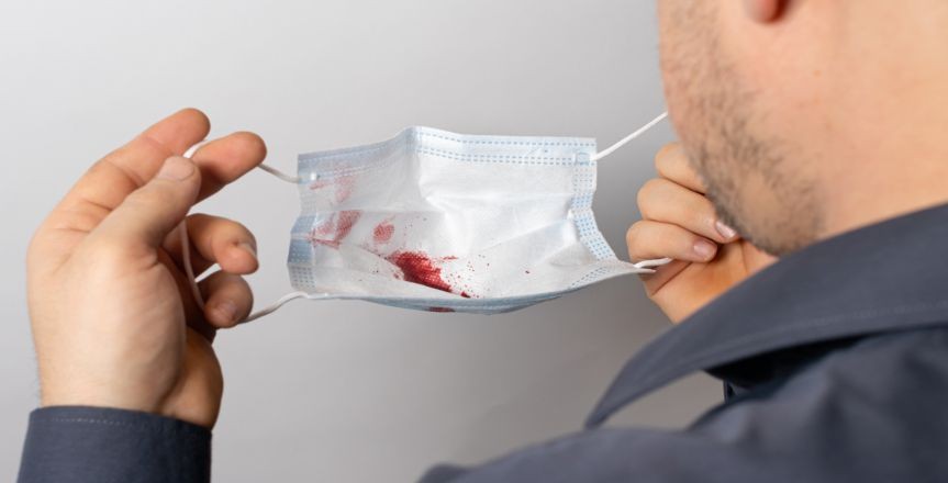
Hemoptysis Versus Hematemesis
Hemoptysis [Coughing up blood]Hemoptysis is when a person is coughing up blood from the lower regions of the respiratory tract usually indicating a problem related to the respiratory or cardiovascular system. There are several reasons why a person may cough up blood in respiratory conditions such as pneumonia, tuberculosis, bronchiectasis, and cystic fibrosis. Simply coughing too hard with too much pressure, and too much can lead to some bleeding due to microscopic vessel rupture which can happen if a person has a respiratory illness. A pulmonary embolism, lung cancer, and even cardiovascular disease can also lead to hemoptysis. Bleeding may be mild or in extreme cases, severe. It is when hemorrhage is uncontrolled that the risk of death is increased and treatment to stop the bleeding becomes urgent and necessary. Hematemesis [ vomiting up of blood]Hematemesis is when a person vomits blood, usually indicating bleeding in the digestive system. This may be confused with hemoptysis which is bleeding from a different system. The most common causes of vomiting blood are problems in the gastrointestinal system. A peptic ulcer, gastritis, and malignant stomach tumors all are causes of hematemesis. Gastric varices /esophageal varices are when blood vessels dilate/swell up and burst in the stomach and esophagus, usually occurring with portal hypertension/liver failure. A Mallory–Weiss tear is a tear in part of the esophagus due to the excessive force of vomiting or even coughing, which may result in bleeding. Alcohol and NSAID drugs badly inflame the stomach and have been implicated in causing hematemesis. Bleeding can be dangerous leading to anemia but most importantly, shock due to blood loss, which could lead to death. Hematemesis can be dangerous and often the doctor needs to perform an endoscopy to locate the source of bleeding and stop it. Bleeding is stopped is usually by sclerotherapy or endoscopic band ligation. The sclerotherapy procedure is when a substance is injected into a blood vessel that causes the blood to clot. Band ligation is when an elastic band is put around a bleeding vein. Difference Between Hemoptysis and Hematemesis - 1) There is usually a tingling sensation in the throat in hemoptysis while in hematemesis the patient will usually complain of nausea and upset stomach. 2) The blood is usually frothy and bright red in hemoptysis while it is dark red in hematemesis, non-frothy and food particles may also be present at the same time. 3) Blood in hematemesis will give an acidic pH when tested with litmus paper whereas that in hemoptysis will be neutral to alkaline. 4) Stools will be almost always positive for occult blood in hematemesis while it is usually negative in case of hemoptysis. But it can also be positive at times if the patient has swallowed his sputum.
More detailsPublished - Tue, 11 Oct 2022
Created by - Rigomo Team
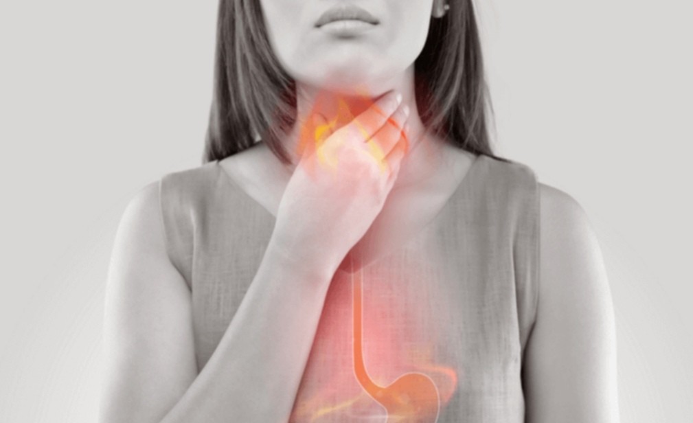
Gastroesophageal reflux disease (GERD): Symptoms, Causes &Treatment
Gastroesophageal reflux disease (GERD) is brought on by stomach acid that frequently rushes back into the tube that connects your mouth and stomach (esophagus). This backwash may irritate the lining of your esophagus (acid reflux). INCIDENCE: 33% of people have symptoms at least once a month, while 10% of people have symptoms every day. 70% of pregnant women experience reflux. PATHOGENESIS: Lower esophageal sphincter pressure loss is what leads to reflux. Pregnancy and medications may influence the tone of the lower esophageal sphincter. Relaxation may be aided by physical disorders like mid-body obesity or a hiatal hernia.CLINICAL FEATURES: The reflux of acidic gastric contents into the esophagus causes a retrosternal, burning pain that may radiate to the neck or jaw. More than 50% of “chest pain” seen in the ED may be reflux.a) The pain is often exacerbated by activities that overcome the lower esophageal sphincter resting pressure such as bending over, lying down, or consuming large meals.b) Uncomplicated GERD is classically relieved within minutes by sitting up and drinking liquids or taking antacids.DIFFERENTIAL DIAGNOSES: Pain from GERD can be indistinguishable from chest pain of cardiac origin; therefore, a cardiac cause must be ruled out. The resolution of symptoms after a trial of oral antacids in the ED does not necessarily exclude a cardiac cause because many patients have both cardiac disease and GERD.EVALUATION is primarily done on an outpatient basis and may include an upper gastrointestinal series, ambulatory acid reflux testing, endoscopy, or manometry to look for spasms as the cause of pain.THERAPY entails the following supportive measures:a) Elevating the head of the bed 6 inches (or 30 degrees) and avoiding recumbency within 3 hours of eating.b) Eating smaller meals and avoiding fatty foods, chocolate, caffeine, peppermint, or alcoholc) Smoking cessationd) Weight reduction (in obese patients)e) Over-the-counter antacids, histamine-2 antagonists (e.g., ranitidine, cimetidine), or other agents such as proton pump inhibitors (PPIs; e.g., omeprazole, pantoprazole) are available for short-term use.DISPOSITION: Admission is recommended for patients at risk for coronary disease. If a cardiac etiology can be ruled out, the patient may be safely released and undergo additional outpatient evaluation.
More detailsPublished - Wed, 12 Oct 2022

Created by - Rigomo Team
Major causes of HEMOPTYSIS
In the hospital setting, the foremost common causes of hemoptysis symptoms are acute bronchitis, pneumonia, T.B., and carcinoma. The diagnosis is made and the underlying etiology of hemoptysis is identified · INFECTIONInfection is the commonest reason for hemoptysis, accounting for 60 to 70% of cases. The infection causes superficial membrane inflammation and edema which may cause the rupture of the superficial blood vessels. The invasive microorganism (e.g., staph aureus, Pseudomonas aeruginosa) or fungi (e.g., Aspergillus species) are the foremost common infectious causes of hemoptysis. Viral infections can also cause severe hemoptysis. HIV infection predisposes patients to many conditions which can lead to coughing up blood. · CANCERPrimary respiratory organ cancers account for 23% of cases of hemoptysis in the United States. Bronchogenic carcinoma is the common carcinoma accountable for hemoptysis in 5 to 54% of all cases. Bleeding from malignant or benign tumors can occur secondary to superficial membrane invasion, erosion into blood vessels, or extensive tracheal involvement.Breast, renal, and colon cancers have a predilection for respiratory organ metastasis but the pathological process of lung metastasis seldom leads to hemoptysis. Certain lesions might catch a secondary infection, leading to hemoptysis. · IDIOPATHICIdiopathic is a diagnosis of exclusion. In 7 to 34% of patients with hemoptysis, no distinctive cause is found after careful analysis. The prognosis for idiopathic hemoptysis is nice, and also the majority of patients have a resolution of this symptom within 6 months of diagnosis. However, results from one study found that an increased incidence of carcinoma in smokers older than forty years with this symptom must prompt these patients might warrant immediate quitting. HEMOPTYSIS IN kids The major reason for this symptom in kids is respiratory tract infection. The second commonest cause is foreign body aspiration, with most cases occurring in kids younger than four years. Another necessary cause is bronchiectasis, which frequently is secondary to mucoviscidosis. Primary phthisis may be a rare cause. Though uncommon, trauma is another possible cause. Blunt-force trauma might end in this symptom secondary to contusion and hemorrhage. Injury caused by suffocation, deliberate or accidental could also lead to hemoptysis.
More detailsPublished - Wed, 12 Oct 2022
Search
Popular categories
Health and Wellness
231Skill Development
7Technology
5Community Impact
2Success story
2Creativity
1Latest blogs

DeepSchool: The Story of an Idea That Refused to Sit Still
Tue, 02 Dec 2025

Transforming Emergency Care: The Story Behind Rigomo's Revolutionary PPMMP Course
Sun, 12 May 2024

Empowering Rural Healthcare: How Pogiko's AI is Bridging the Gap in Medical Services
Thu, 25 Apr 2024
Write a public review