
Created by - Rigomo Team
Hypocalcemia: Causes, Clinical features & Treatment
Ionized calcium level below 2.0 mEq/L or a total serum level below 8.5 mg/dL.Causes of hypocalcemia: Shock, sepsis, renal failure, pancreatitis, hypomagnesemia, alkalosis, decreased serum albumin, hypoparathyroidism (idiopathic or as a result of irradiation or surgery), pseudo-hypoparathyroidism, osteoblastic metastasis, malabsorption, and excess phosphates.CLINICAL FEATURES1. Symptoms: Circumoral and distal extremity paresthesias, irritability, weakness, fatigue, muscle cramps, and seizures2. Physical examination findings: Hyperreflexia, carpopedal spasm, tetany, laryngospasm, Trousseau sign (carpopedal spasm after arterial occlusion of the arm for 3 minutes), and Chvostek sign (contraction of the facial muscles after percussion over the facial nerve)EVALUATION1. Laboratory studies should include serum albumin, calcium, magnesium, phosphate, BUN, and creatinine levels; liver studies; amylase and lipase levels; ionized calcium levels; a serum electrolyte panel; and a CBC.2. Electrocardiography: ECG findings may include a prolonged QT interval, sinus bradycardia, complete heart block, ventricular arrhythmias, and ventricular fibrillation.3. Radiology: When hypocalcemia occurs in the context of osteomalacia, radiographic findings can include craniotabes, frontal skull bossing, rachitic rosary ribs, a widened rib cage (Harrison groove), bowed legs, demineralization, and thinning of the cortical bone.THERAPY1. Acutely symptomatic hypocalcemia: Administer 10 mL of 10% calcium gluconate infused intravenously over 10 to 15 minutes, followed by a maintenance infusion of 1 to 2 mg/kg/hour over 6 to 12 hours.2. Asymptomatic: Oral therapy with elemental calcium (with or without vitamin D) may be all that is required. The rapid intravenous administration of calcium to asymptomatic patients with mild to moderate hypocalcemia is contraindicated because doing so can cause severe cardiovascular, neuromuscular, or renal complications.DISPOSITION1. Admission: Patients with symptomatic hypocalcemia who require intravenous replacement therapy must be admitted to the hospital. These patients should be placed on continuous cardiac monitoring, and serial serum calcium levels should be obtained.2. Discharge: Asymptomatic patients may be discharged with appropriate follow-up.
More detailsPublished - Wed, 12 Oct 2022

Created by - Rigomo Team
Treatment Plan for Haemoptysis
Mild cases do not need admissions, They are resolved by the use of cough suppressants. Moderate-to-severe haemoptysis, though a rare event, need evaluation to find out the cause of bleeding. o Antibiotics are given for pneumonia and TB. Tuberculosis has been known as the foremost common causes of serious haemoptysis in recent empiric studies throughout the globe.o Steroids are prescribed for the inflammatory condition under the supervisionBleeding can be stopped by embolizing (blocking) a bleeding artery/through a bronchoscopy after evaluating the patient’s underlying condition.Surgical intervention is needed for cancer Haemoptysis Considerationso It is reported that 5–14% of patients presenting with haemoptysis can have severe haemoptysis or additionally referred to as huge haemoptysis, which has been variably outlined primarily based upon criteria like the amount per hour of blood, the full volume of blood per 24 h, or the presence of abnormal blood gas values or hemodynamic instability.o Normally, blood loss > 100 mL/h or total volumes > 500 mL in 24 h is thought to be grave haemoptysis. o Depending upon the patient’s underlying respiratory organ condition, smaller volumes (50 mL) of haemoptysis are also grave.
More detailsPublished - Thu, 13 Oct 2022

Created by - Rigomo Team
Hypercalcemia: Causes, Symptoms & Treatment
Total calcium level exceeding 10.5 mg/dL or an ionized calcium level exceeding 2.7mEq/LCAUSES1. Endocrine causes: Primary hyperparathyroidism, hyperthyroidism, pheochromocytoma, adrenal insufficiency, and acromegaly2. Malignancies: Squamous cell carcinoma of the lung, breast cancer, kidney cancer, myeloma, and leukemia3. Granulomatous disorders: Sarcoidosis, tuberculosis, histoplasmosis, and coccidioidomycosis4. Medications: Excessive vitamin D or A intake, thiazides, lithium, and hormonal therapy for breast cancer can cause hypercalcemia.5. Miscellaneous: Immobilization, Paget disease, dehydration, excess calcium ingestion, and milk-alkali syndromeCLINICAL FEATURES1. Signs and symptoms: Weakness, depression, confusion, lethargy, personality changes, nausea, vomiting, anorexia, constipation, headache, and abdominal pain2. Physical examination findings: Dehydration, decreased motor strength, decreased mental status, ataxia, hyporeflexia, fractures, hypertension, weight loss, renal insufficiency, and cardiac arrestEVALUATION1. Laboratory studies should include ionized calcium levels; serum calcium, protein, phosphate, magnesium, BUN, creatinine, glucose, amylase, and lipase levels; a serum electrolyte panel; and a CBC.2. Electrocardiography: ECG abnormalities include shortening of the QT interval, widening of T waves, bradyarrhythmias, bundle branch blocks, and second-degree and complete heart block.THERAPYTreatment is required for symptomatic patients with calcium levels greater than 12mg/dL who are unable to maintain a good fluid intake or have abnormal renal function.1. Fluid replacement: Because patients with hypercalcemia are usually dehydrated, the initial and safest treatment is the restoration of volume with large amounts of saline (5 to 10 L of normal saline in the first 24 hours).2. Pharmacologic therapya) Furosemide (1 to 3 mg/kg) can be administered intravenously to enhance urinary output and increase renal excretion of calcium.b) Calcitonin (2 to 4 IU/kg intramuscularly every 12 hours) diminishes calcium levels, usually within 12 hours. Calcitonin is useful for the initial treatment of symptomatic hypercalcemia greater than 14 mg/dL.c) Bisphosphonates are used for the longer-term management of hypercalcemia related to bone resorption.3. Dialysis: Patients with severe symptoms and patients with cardiac or renal diseaseDISPOSITIONPatients with a calcium level greater than 12 mg/dL, symptoms or abnormal renal function require admission for continuous cardiac monitoring and serial calcium levels.
More detailsPublished - Thu, 13 Oct 2022

Created by - Rigomo Team
All you want to know about COPD
Chronic Obstructive Pulmonary Disease (COPD) refers to a group of diseases that has bronchitis and Emphysema. Over time, COPD makes it tougher to breathe. You can’t reverse the damage to the lungs, however, lifestyle changes and medication will assist you to manage the symptoms.o Chronic bronchitis irritates your bronchial tubes, which carry air to and from your lungs. In response to irritation, the tubes swell and secretion (phlegm or “snot”) builds up on the lining. The build-up narrows the passage, making it difficult to push air into and out of your lungs.Small, hair-like structures called cilia unremarkably move secretion out of your airways. however, the irritation and inflammation from bronchitis and/or smoking damage them. The broken cilia can’t facilitate clear secretion.o EmphysemaEmphysema is the breakdown of the walls of the little air sacs (alveoli) at the tip of the bronchial tubes, within the “bottom” of your respiratory organ. Your respiratory organ is like a tree. The trunk is the cartilaginous tube or “trachea,” that branches into the “bronchi,” which further terminate into the air sacs or “alveoli.”The air sacs play a vital role in transferring oxygen into your blood and carbon dioxide out. The damage caused by respiratory disorder destroys the walls of the air sacs, making it exhausting to take a full breath.What are the causes of COPD?The primary cause of COPD is smoking. however, not all smokers develop the sickness. you'll be at higher risk if you:o Are over the age of sixty-five.o Have been exposed to pollution for a long time.o Have occupational exposure to toxic chemicals or fumes.o Have alpha-1 antitrypsin deficiency (AAT), a genetic risk issue for COPD.o Had a history of repeated infections throughout childhood. What are the signs and symptoms of Chronic Obstructive Pulmonary Disease?o Cough with copious secretions that persists for a long time.o Difficulty taking a deep breath.o Shortness of breath with gentle exercise (like walking or climbing stairs).o Shortness of breath playing regular daily activities.o Wheezing. How is COPD diagnosed?To assess your lungs and overall health, your healthcare provider will medical history, perform a physical examination, and order some tests, like PULMONARY FUNCTION TESTS.Medical historyTo diagnose COPD, your doctor can ask queries like:o Do you smoke?o Have you had long-run exposure to dust or air pollutants?o Do any members of your family have COPD?o Do you get breathless with exercise? Or even while resting?o Have you had a cough for a very long time?o Do you cough up phlegm?Physical ExaminationTo help with the diagnosis, your doctor can do a physical examination that includes:o Listening to your lungs and heart.o Checking your vital sign and pulse.o Examining your nose and throat.TestsProviders use an easy check known as SPIROMETRY to ascertain how well your lungs work. For this check, you blow air into a tube connected to a machine. This respiratory organ performs check measures what quantity of air you'll inhale in a breath and how quickly you'll exhale.Your doctor might also wish to run a couple of other tests, such as:o Pulse oximetry: This check measures the oxygen levels in your blood.o Arterial blood gases (ABGs): These tests check the oxygen and carbon dioxide levels.o Electrocardiogram (ECG or EKG): This checks on the heart and rules out cardiovascular disease as a reason for shortness of breath.o Chest X-ray or chest CT scan: Imaging tests seek for changes that COPD causes. o Exercise testing: Your doctor uses this to work out if the oxygen level in your blood drops after you exercise.
More detailsPublished - Fri, 14 Oct 2022
Created by - Rigomo Team
Diabetic Ketoacidosis (DKA): Etiology, Clinical features & Treatment
In insulin-dependent patients, diabetic ketoacidosis is characterized by hyperglycemia, ketonemia, and acidosis.ETIOLOGYDiabetic ketoacidosis is caused by a relative or absolute deficiency of insulin and increased levels of stress hormones (e.g., catecholamines, cortisol, growth hormone). Insulin deficiency leads to lipolysis, which in turn leads to the production of ketone bodies, resulting in acidosis.PRECIPITATING FACTORS include lack of insulin, infection, injuries, emotional stress, alcohol use, myocardial infarction, and cerebrovascular accident.CLINICAL FEATURES1. Symptoms: Nausea, vomiting, abdominal pain, polyuria and polydipsia, and altered mental status.2. Physical examination findingsa) Kussmaul respirations (i.e., rapid, deep breathing) may be noted, and the breath often smells like acetone.b) Dehydration may be reflected by hypotension, reflex tachycardia, dry skin, and dry mucous membranes.DIFFERENTIAL DIAGNOSESHypoglycemia, nonketotic hyperosmolar coma, isopropyl alcohol ingestion, alcoholic ketoacidosis, lactic acidosis, uremia, toxin ingestion, and starvation ketosis must be ruled out.EVALUATIONLaboratory studies:1. A provisional diagnosis can be obtained via blood gas and bedside glucose determinations.a) There will be signs of hyperglycemia, which is indicated by a blood glucose level of at least 300 mg/dL.b) Metabolic acidosis is demonstrated by a serum bicarbonate concentration of less than 15 mEq/L and a pH of less than 7.3.2. A serum biochemical profile (including electrolytes, blood urea nitrogen [BUN], and creatinine levels), urinalysis, and ketone levels confirm the diagnosis.a) Ketonemia results from -hydroxybutyrate and acetoacetate. Qualitative tests (e.g., the nitroprusside test) detect acetoacetate but not β-hydroxybutyrate.b) Depending on the patient's level of hydration, electrolyte imbalances may exist.3. Other studies, such as a complete blood count (CBC), a chest radiograph, or an electrocardiogram (ECG), may be indicated to identify precipitating causes.THERAPY1. Normal saline should be administered at an initial rate of 1 L/hour for the first 2 to 3 hours. The average fluid deficit is 5 to 10 L. Clinical response and urine output are the best indicators of fluid status.2. Insulin is administered intravenously as a continuous infusion using a low-dose technique (i.e., 5 to 10 U/hour) until the ketonemia and acidosis have resolved.3. Potassium (20 mEq/L) should be added to the intravenous fluids during early therapy to correct the profound potassium deficiency associated with diabetic ketoacidosis. During the first day of treatment, the patient usually requires 100 to 200 mEq of potassium.4. Phosphate: Whether phosphate replenishment is required is debatable. Phosphate is given either orally or intravenously if the patient’s serum phosphate level decreases to below 1 mg/dL.DISPOSITIONMost patients with diabetic ketoacidosis need to be admitted, often to an intensive care unit (ICU). In patients with mild diabetic ketoacidosis, the ketoacidosis may resolve in the emergency department. These patients should be placed under observation until any underlying precipitating causes can be ruled out.
More detailsPublished - Sat, 15 Oct 2022

Created by - Rigomo Team
Is It Necessary To Change Diet With Changing Weather?
On some days, it's summer, and then comes a relief with the monsoon season. So, all in all, the weather is continually changing and it's very troublesome to keep up your diet in line with the season. However, to stay your health in restraint, it's vital to eat the proper healthy food as per the body’s needs in a particular season and keep yourself stay robust while facing erratic weather changes. The food we eat plays a crucial role in serving our bodies handle the changes in the weather, especially if you've got the tendency towards falling sick with changing temperatures. More often people get a common cold and sore throat with seasonal changes. It may not appear serious but with the similarity of symptoms with COVID, it creates panic and may encourage people to have proper healthy food to build a robust immune system and ability to withstand harsh changes and help serve the body to regulate the changes and adaptation to the weather.Some of the food and dietary habits help assist the body in adapting to seasonal change:1. Vitamin C: Much evidence supports the role of vitamin C in improving immunity. Citrus fruits and vegetables such as vegetables like broccoli, cauliflower, and kimchi are an important part of a healthy diet. These vegetables add flavor, help increases digestive health, and may help lose weight. 2. Increase your liquid intake: It helps with digestion, removes toxins, and additionally regulates temperature. However different liquids also can be equally effective in maintaining a healthy body such as fruit juices, tomato soup with herbs, and lentil soups. Aside from that, make sure to incorporate healthy drinks like turmeric milk, and homemade curries with spices, ginger barley, or lemon broth into your diet. 3. Avoid raw foods: Even if raw foods are extremely nourishing, they're very hard on our digestive system, which makes it troublesome for our bodies to digest them. In addition, within the monsoon seasons, these raw veggies may attract heaps of bacteria that may additionally affect our health. However, as mentioned earlier exposure to heat will affect the nutrition of some foods. However, it's great to steam your food before consuming it.4. Eat seasonal crop: currently, the catch here is that it's troublesome to decide what seasonal crop to eat for example methi, which is actually a winter crop is currently out there throughout the year. However, check that to eat all the fruits and vegetables that are out there at your native market. Fruits like pineapples and mangoes are typically available during a specific season. However, Millet, sorghum, and maize are stronger crops that don't seem to be simply affected by weather changes.5. Sour food: Sour foods work wonders for your gut. Digestion is improved with the consumption of foods like kombucha, kanji, and yogurt in your daily diet. With erratic weather changing regularly healthy sterile food guarantee stabilizing the physiological condition of the body.
More detailsPublished - Sat, 15 Oct 2022
Created by - Rigomo Team
Ectopic Pregnancy: Risk factors, Clinical Features & Differential Diagnoses
Ectopic pregnancy is the development of a fertilized ovum outside of the uterine cavity (e.g., in the fallopian tube, ovary, cervix, or abdominal cavity). The ectopic site can rarely sustain the pregnancy beyond several weeks, at which time the implantation site ruptures.PATHOGENESISThe fertilized ovum implants at the ectopic site, stimulating a persistent corpus luteum. The resultant elevated estrogen levels stimulate endometrial growth, and progesterone maintains this lining for uterine implantation that never arrives. The ectopic pregnancy continues to proliferate until it outgrows its blood supply and involutes or ruptures.RISK FACTORS are generally related to tubal dysfunction or injury and include:a) Tubal anomalies (e.g., hypoplasia, diverticula)b) Salpingitis (characterized by inflammation, scarring, and lumen narrowing)c) Tubal adhesions (e.g., from infection or endometriosis)c) Previous tubal surgery (e.g., salpingostomy, tubal ligation)d) Intrauterine device usee) Previous ectopic pregnancyCLINICAL FEATURESThe “classic triad” of a missed period, abdominal pain, and a palpable mass on examination are present in fewer than 30% of patients. Important historical and clinical findings include the following:1. Menstrual history: A history of amenorrhea or a late period is common in patients with ectopic pregnancy. Only 10% of patients describe a normal last menstrual period.2. Abdominal pain or tenderness: Ninety percent of patients complain of abdominal or pelvic pain.a) The pain usually begins as colicky and diffuse (as a result of ectopic distention and inflammation) and later becomes localized (as a result of inflammation of the adjacent abdominal wall and local bleeding).b) Peritoneal symptoms may be noticed if the bleeding causes diffuse peritoneal irritation. With severe bleeding and peritonitis, the abdomen will be rigid, distended, and tender.3. Cervical motion tenderness [It is cervical excitation that is observed while performing the pelvic examination and is a classical sign suggestive of pelvic pathology] or adnexal tenderness [pain on touch, in the area of a woman's uterus ] is highly suggestive of a pathologic process. Adnexal tenderness is a likely finding in ectopic.4. Vaginal bleeding: Fifty percent to 90% of patients will note abnormal bleeding, ranging from spotting to heavy flow with large clots.5. Uterine enlargement: In pregnancy, the uterus softens and grows in response to hormonal stimulation regardless of the site of conceptus implantation. One cannot assume the pregnancy is intrauterine based on uterine size.6. Palpable mass: Experienced examiners may note a unilateral or cul-de-sac mass, although the absence of such a mass does not rule out an ectopic pregnancy.7. Volume depletion: Tachycardia, orthostatic hypotension, near-syncope, abdominal pain, and a positive pregnancy test in an otherwise healthy woman are indicative of a ruptured ectopic pregnancy until proven otherwise.DIFFERENTIAL DIAGNOSES — Miscarriage— Ovarian cyst— Vaginitis— Cervicitis— Salpingitis— PID— Combined pregnancy (i.e., intrauterine and ectopic, may be seen in patients taking infertility medications)— Normal intrauterine pregnancy— Appendicitis— Urinary tract infection— Acute nephrolithiasis— Enteritis— Diverticulitis
More detailsPublished - Sat, 15 Oct 2022

Created by - Rigomo Team
Treatment approach for COPD
Chronic Obstructive Pulmonary Disease, or COPD, is the umbrella term for a bunch of respiratory organ diseases that include bronchitis and Emphysema. A COPD diagnosis means that you have either one of those lung-damaging diseases.Although there’s no cure for this condition, and also lung injury can’t be reversed, however, you can slow its progression and manage symptoms by taking the correct steps and working closely together with your doctor.Prevention -The best way to prevent COPD is never to start smoking or quit smoking to stop the progression of the disease, in case you smoke. Smoking is accountable for up to eight out of ten COPD-related deaths, and 38 % of adults diagnosed with COPD report current smoking, as per CDC reportsSmoking affects your airways, which causes irritation and swelling.For advanced stages, many effective therapies may relieve symptoms, slow the decline of respiratory organ performance, cut back your risk of complications and exacerbations, and improve your ability to steer an energetic life.Common treatments for COPD include:Bronchodilator medication relaxes the muscles around the airways, which helps to stay them open and makes breathing easier. Most bronchodilators are delivered through a dispenser or are nebulized straight into your lungs through the breath.o Bronchodilators embody beta-agonists and anticholinergics which may be either short-acting or long. Short-acting bronchodilators offer immediate relief of symptoms; however, the effect may wear off in a very few hours. Long-acting bronchodilators give relief for several hours, however, could take longer to kick in. o Corticosteroids (Steroids) These medicines help reduce swelling and mucous production in the airways, making it easier to breathe. Steroids can be given as inhalers, or may also be taken as a pill
More detailsPublished - Mon, 17 Oct 2022
Created by - Rigomo Team
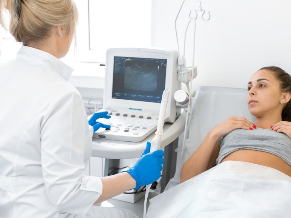
Ectopic Pregnancy: Evaluation & Treatment
EVALUATIONThe incidence of detected ectopic pregnancies has increased fivefold over the last 20 years due to increasingly sensitive screening aids.1. Stabilization: It is important to establish the airway, respiratory, and cardiovascular status immediately before making a diagnosis. Two large-bore peripheral lines are secured for fluid and blood resuscitation.2. Laboratory studiesa) Urine pregnancy test: A urine sample can provide a quick qualitative β–human chorionic gonadotropin (β-hCG) screen and most screens with increasing sensitivity, now at approximately 4 weeks gestational age.b) Blood work: Blood typing and screening, Rh sensitivity, quantitative β-hCG level, and a CBC are sent immediately. Β-hCG levels normally double every 36 to 48 hours. Levels in abnormal pregnancies do not increase as rapidly and may plateau or decline. Thus, if they are available, serial levels are useful in establishing a growth pattern.3. Ultrasonography: A distinct fetal body and fetal cardiac activity can be detected ultrasonographically by 6 weeks gestation (transvaginal) or 7 weeks gestation (transabdominal), making the search easier for the ectopic site at this stage. There must be two distinct echogenic layers (i.e., the decidua vera and the decidua capsularis) surrounding the gestational sac before the diagnosis of intrauterine pregnancy can be made.THERAPY1. General therapy: All Rh-negative mothers are given Rho(D) immune globulin in a single intramuscular dose to prevent potential maternal Rh antibody formation. Can be within 72 hours of the initial bleeding episode2. Specific therapya) Unruptured ectopic pregnancy: These patients are hemodynamically stable. Treatment options include:— Laparoscopic salpingostomy and ectopic excision— Nonsurgical treatment with methotrexate, leucovorin, or mifepristone— Salpingocentesis (i.e., transvaginal needle aspiration of the gestational sac, potentially with direct injection of methotrexate)b) Ectopic pregnancy without cardiac activity: Patients with a serial decline of β-hCG levels and no evidence of erosion or rupture on repeat sonography may be managed nonoperatively by the obstetrician. Medications may be used to induce expulsion. Laparoscopic intervention is performed if there is any sign of erosion or rupture.c) Ruptured ectopic pregnancy: These patients may be hemodynamically unstable and therefore require aggressive fluid and blood resuscitation and control of bleeding. Laparoscopic ectopic excision via salpingectomy is indicated.DISPOSITION1. A hemodynamically stable patient with a history of abdominal pain, vaginal bleeding, or both; a benign examination; and ultrasound revealing no clear evidence of ectopic or intrauterine pregnancy may be discharged with strong ectopic precaution warnings. These patients should see an obstetrician in 48 hours for a repeat examination and quantitative β -hCG levels. Ultrasound is repeated in 2 to 7 days to search for pregnancy.2. A hemodynamically stable patient with a history of abdominal pain, vaginal bleeding, or both with abdominal or adnexal tenderness on physical examination, and evidence of cul-de-sac fluid accumulation on ultrasound but no clear evidence of intrauterine pregnancy should be admitted for observation, serial abdominal examinations, repeat quantitative β-hCG levels (in 36 to 48 hours), and repeat sonography.3. A hemodynamically stable patient with or without abdominal or adnexal tenderness and sonographic evidence of ectopic pregnancy is admitted.4. A hemodynamically unstable patient with pregnancy by qualitative β-hCG requires aggressive resuscitation en route to the operating room.
More detailsPublished - Tue, 18 Oct 2022
Search
Popular categories
Health and Wellness
231Skill Development
7Technology
5Success story
2Community Impact
2Strategy
1Latest blogs

DeepSchool: The Story of an Idea That Refused to Sit Still
Tue, 02 Dec 2025

Transforming Emergency Care: The Story Behind Rigomo's Revolutionary PPMMP Course
Sun, 12 May 2024

Empowering Rural Healthcare: How Pogiko's AI is Bridging the Gap in Medical Services
Thu, 25 Apr 2024
Write a public review